3413
Dual-echo blip reversed EPI acquisition enables distortion correction in the presence of motion in diffusion-weighted MRI1Boston Children's Hospital and Harvard Medical School, Boston, MA, United States, 2Brigham and Women's Hospital and Harvard Medical School, Boston, MA, United States
Synopsis
Slice-to-volume registration methods have been shown to provide motion robust reconstruction for large and frequent motions. One challenge with motion correction is the changing magnetic field inhomogeneities with different head positions. In this work we implemented a dual-echo blip reversed EPI acquisition and show that this sequence can be used to reduce distortions in large and frequent motions and can improve slice-to-volume registration results.
Introduction
Echo planar imaging (EPI) is inherently susceptible to distortions arising from local magnetic field inhomogeneities. Field maps calculated from gradient echo images or EPI images with opposing phase encoding directions can be acquired before a scan to correct for distortion. Unfortunately when imaging uncooperative patients, such as young children, infants, and fetuses, subject motion interferes with distortion correction using static field mapping as the field changes with different patient positions. Here, in this work, we implemented an alternative approach1, where we acquire a blip-reversed EPI readout using multiple echoes for each slice. We use these echoes, acquired 30-50ms apart effectively freezing the motion, to correct for distortions on each slice and then use a slice-to-volume registration (SVR) strategy2 that has been shown to produce robust reconstructions even under large and frequent patient motion.Methods
A standard diffusion weighted EPI sequence was modified with a 1800 RF pulse and an additional echo with an opposing ky-blip polarity compared to the first echo. We used a standard 30-direction single shell diffusion weighting with a b-value of 1000 and three B0 images. After both echoes with opposing phase encodes were reconstructed, we used a slice-level distortion correction method3 that generates a single distortion free image. The sequence was tested on images acquired for 4 healthy volunteers at 3T (Siemens, Erlangen, Germany). Sequence parameters were TE1=72ms, TE2=108ms, GRAPPA=2, TR=7000ms, 2mm isotropic resolution with 70 slices resulting in a scan time of 6 minutes. Two different sets of experiments were performed on each volunteer:
- In the first set, volunteers were scanned with two identical diffusion acquisitions and they were instructed to move around 10 degrees between each scan. A T2-weighted FSE sequence and a multi-echo 3D GRE sequence were acquired at each position to provide a reference image and field map at each position. We compared the distortion corrected images with T2 images and field maps generated from distortion correction with the reference field map created using the 3D GRE scan.
- In the second set, subjects were instructed to move to a different position using audio commands every minute. An electromagnetic tracker was attached to each subject to measure head motion. The data was processed offline using a slice-to-volume registration method adopted from Marami et. al2. The electromagnetic tracking motion parameters were compared with motion parameters estimated by SVR and raw EPI images and distortion corrected images.
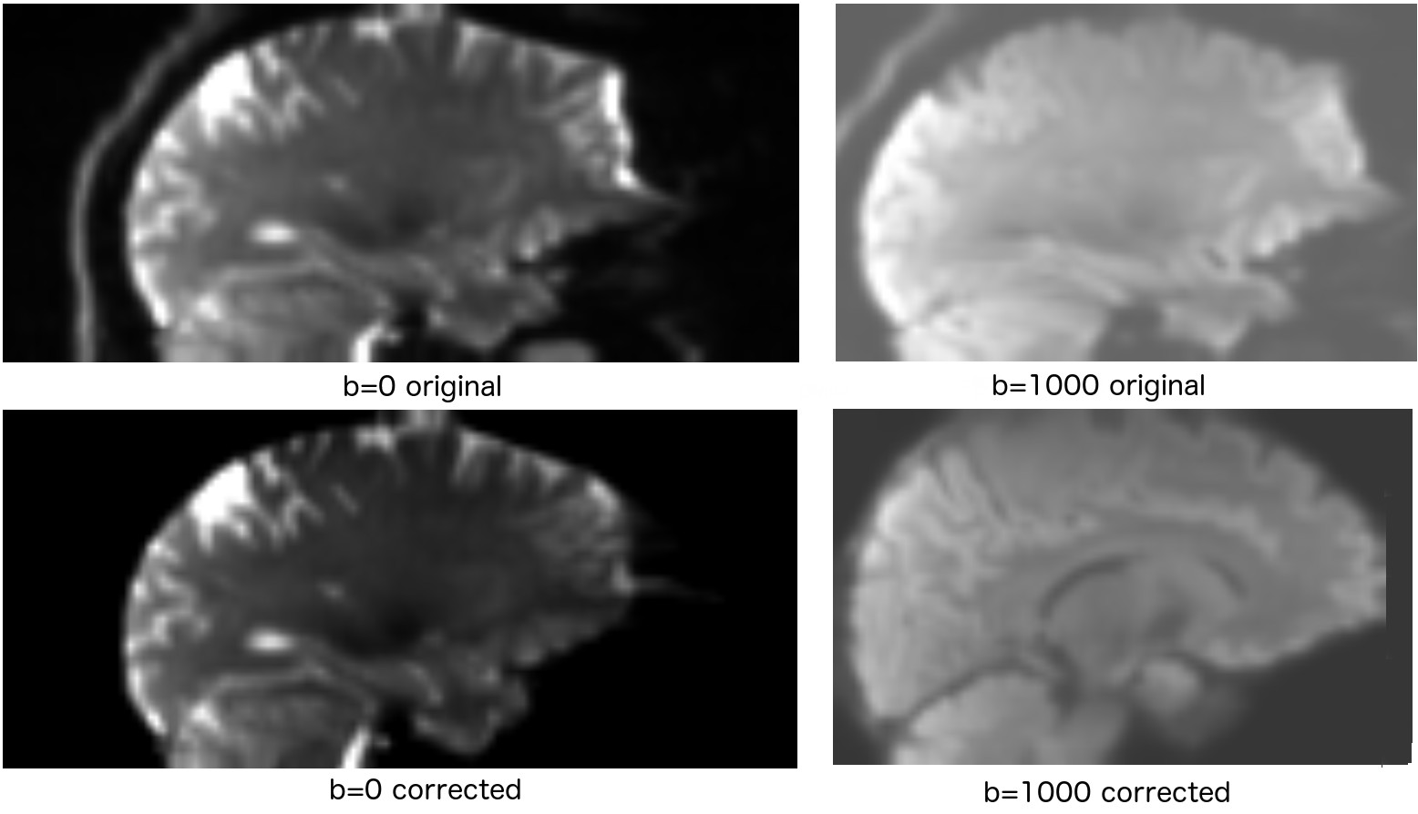
Results
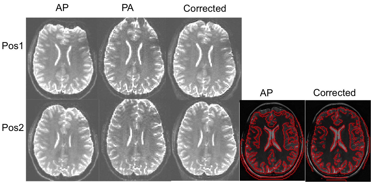
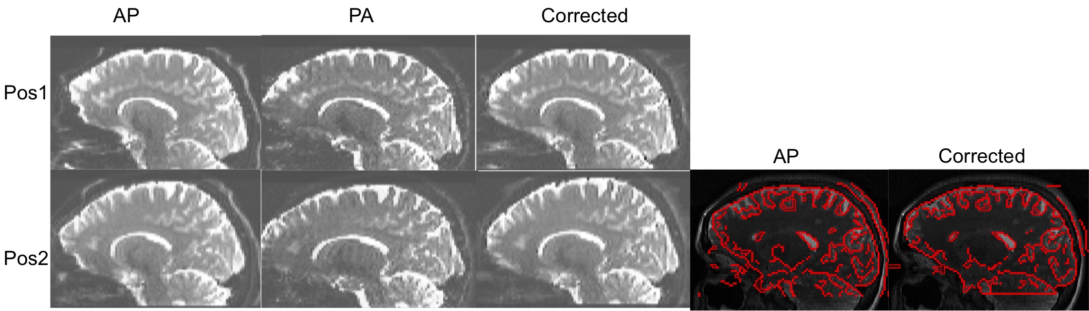
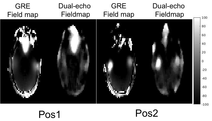
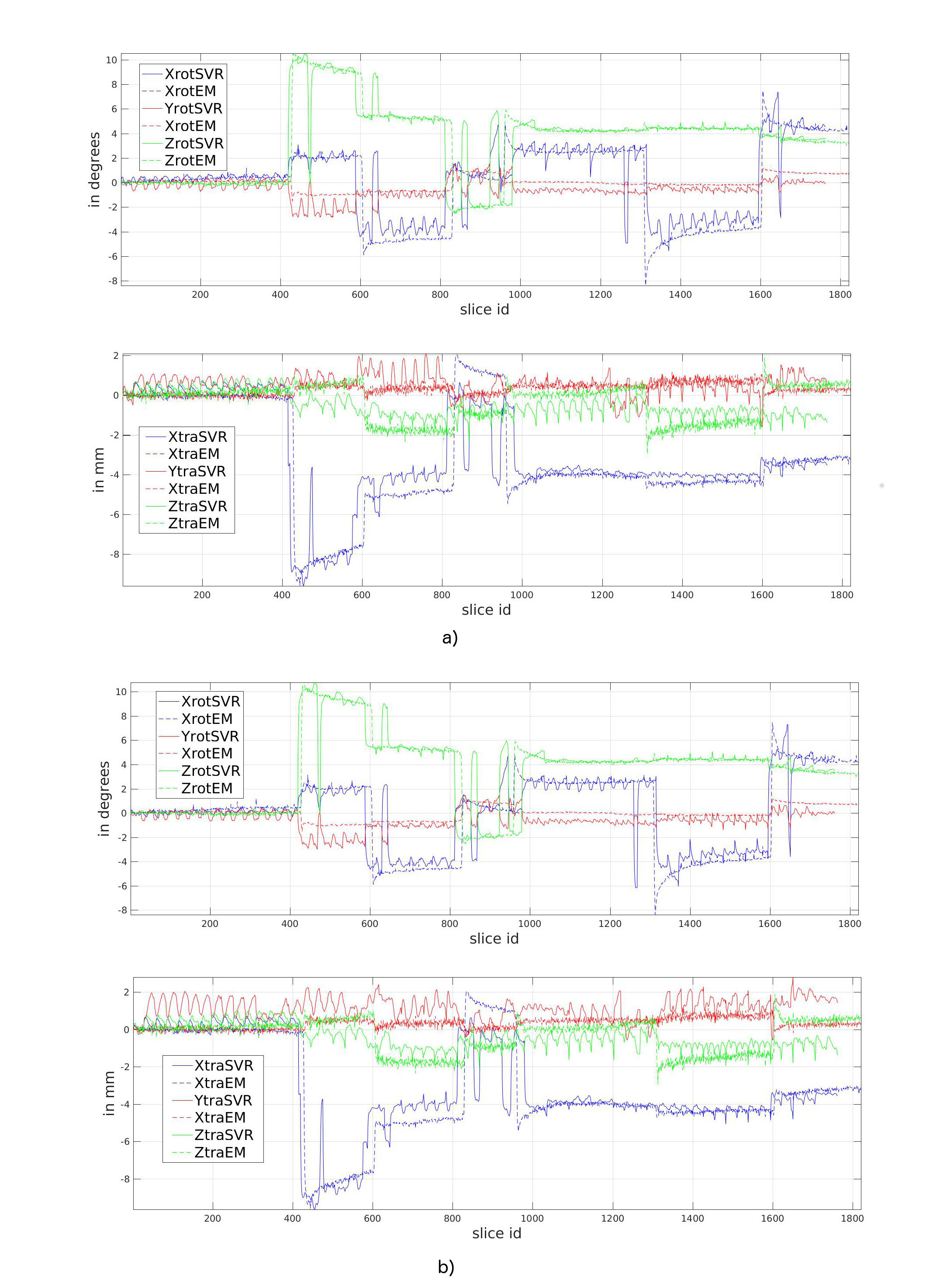
Figure 1 (axial) and 2 (sagittal) show results from a volunteer in two different positions. Even with a large nodding motion (10 degrees) the dual-echo scan was able to compensate for distortions and generated an image that matches well to the structural T2 scan. Figure 3 shows a comparison between field maps generated using the dual echo scan and the reference field maps. The dual echo approach was able to create a similar field map compared to the multi-echo GRE scan at both positions. Figure 4 shows motion parameters reported by EM tracking hardware compared to the SVR results before and after distortion correction. Even with large and frequent motions reported here, SVR was able to generate robust reconstructions. The error in SVR results compared to EM tracking reduced from 0.79 $$$\pm$$$ 1.46mm to 0.62mm $$$\pm$$$ 1.35 for translation and 0.92 $$$\pm$$$ 0.76 degrees to 0.84 $$$\pm$$$ 0.75 degrees for rotation. Figure 5 shows final B0 and mean diffusion image using SVR before and after distortion correction. The distortion from B0 inhomogeneities can be clearly seen in the images generated from uncorrected raw images.Conclusions
Our results show that dual echo acquisitions with blip-reversed phase encoding can be used to generate slice level distortion free images, which is critical for motion-robust slice to volume registration. The distortion corrected images not only resulted in better motion estimates, they also generated a more accurate final diffusion image reconstruction. This method can be used in studies where subject motion is inevitable like body diffusion and fetal brain diffusion studies, and can also be used to reduce the rate of sedation and anesthesia in imaging infants, young children, and uncooperative patients, and in imaging uncooperative patients in research studies.Acknowledgements
This research was supported in part by the following grants: NIH-R01EB019483, NIH-R01NS079788, NIH-R01EB018988, and NIH-R44MH086984.References
1) Gallichan, D., Andersson, J.L., Jenkinson, M., Robson, M.D. and Miller, K.L., 2010. Reducing distortions in diffusion‐weighted echo planar imaging with a dual‐echo blip‐reversed sequence. Magnetic resonance in medicine, 64(2), pp.382-390.
2) Marami, B., Scherrer, B., Afacan, O., Erem, B., Warfield, S.K. and Gholipour, A., 2016. Motion-robust diffusion-weighted brain MRI reconstruction through slice-level registration-based motion tracking. IEEE Trans. Med. Imaging, 35(10), pp.2258-2269.
3) Andersson, J.L., Skare, S. and Ashburner, J., 2003. How to correct susceptibility distortions in spin-echo echo-planar images: application to diffusion tensor imaging. Neuroimage, 20(2), pp.870-888.
Figures