3408
Minimization of Nyquist ghost artifacts for diffusion-weighted single-refocused spin-echo EPI1Brain Imaging Center (BIC), Goethe University Frankfurt, Frankfurt am Main, Germany
Synopsis
In diffusion-weighted (DW) imaging with EPI readout, Nyquist ghost (NG) artifacts might be aggravated due to higher order eddy currents, especially when using monopolar DW gradients in single-refocused spin-echo EPI (srSE-EPI). Both linear and point-by-point phase corrections were tested on DW-srSE-EPI and, for comparison, also on DW twice-refocused spin-echo EPI (trSE-EPI) with intrinsic eddy-current compensation. Both phase correction methods performed equally well for DW-trSE-EPI. However, for DW-srSE-EPI with high b-values, the linear phase correction failed to fully correct NG artifacts. In contrast, point-by-point phase correction yielded considerably better results. This was confirmed in vitro and in vivo.
Introduction
Echo planar imaging (EPI) may suffer from Nyquist ghost (NG) artifacts, due to phase differences between odd and even echoes inside the echo train. They appear as a ghost of the main image, shifted by half the field of view (FOV) along the phase encoding direction. Such NG artifacts are generally corrected by linear [1] or point-by-point [2] phase correction methods, requiring reference echoes that are acquired without phase blips prior to the image acquisition. These reference echoes are used to calculate the phase inconsistency between odd and even echoes. In diffusion-weighted imaging (DWI), an EPI readout is commonly used and images may suffer from even stronger NG artifacts due to higher order eddy currents induced by monopolar diffusion weighted gradients (DWG) in single-refocused spin-echo EPI (DW-srSE-EPI) sequences. The purpose of this study was to determine which phase correction method is best suited for removing the NG artifacts in DWI.Methods
MRI experiments were performed on a 3T whole-body scanner (MAGNETOM Prisma, Siemens Healthineers, Erlangen, Germany), using a body TX and a 64-channel phased-array head/neck RX coil. All protocols were first tested in vitro on an agarose gel phantom, then in vivo on four healthy volunteers. Written informed consent was obtained from all participants before scanning. For each phase correction method (linear, point-by-point), DW data sets were acquired via DW SE-EPI sequences with 30 different DWG directions at a b-value of 500 s/mm2 (in vitro) or 1000 s/mm2 (in vivo). DWG directions were distributed symmetrically, using full-sphere sampling. For the phantom experiments, the diffusion-weighted twice-refocused spin-echo EPI (DW-trSE-EPI) with intrinsic eddy-current compensation (ECC) [3] and the optimized DW-srSE-EPI sequence [4, 5] were used with identical parameters except for TE: in-plane resolution=2×2 mm2, FOV=192×192 mm2, matrix-size=96×96, number of interleaved axial slices=72, slice-thickness=2 mm, no inter-slice gap, echo-spacing time=0.68 ms, bandwidth=1680 Hz/pixel, partial Fourier of 75%, GRAPPA acceleration factor=2. For the phantom experiments, TR=7500 ms, TE=58/70 ms (srSE/trSE) and DWG amplitude (Gmax)=38 mT/m were used. In vivo, TR/TE=6300/55 ms and Gmax=64 mT/m were used in only DW-srSE-EPI sequence.
Online NG corrections were performed using the linear and the point-by-point approach separately. For the linear method, spatially constant and linear phase errors are obtained by a linear regression along the readout direction and subsequently removed from the EPI dataset [1]. Point-by-point phase correction does not perform a linear regression, using for each pixel along the readout direction the locally calculated correction factor [2].
In order to obtain a reference without diffusion weighting and to correct for geometrical distortions induced by static magnetic field inhomogeneities, four reference images at b=0 were acquired with positive and negative phase encoding gradients. To compute fractional anisotropy (FA) maps and principal eigenvectors, preprocessing and subsequent data analysis were performed as explained in the literature [4, 5].
Results
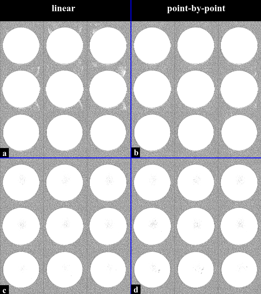
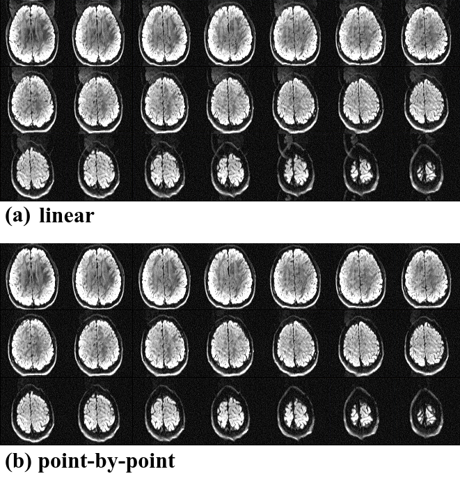
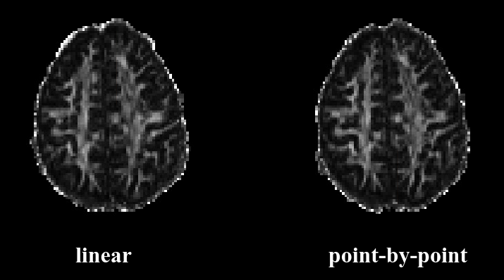
Figure 1 shows the phantom results for both phase correction methods and both DWI sequences, applying linear (left) and point-by-point (right) phase correction to datasets acquired with optimized DW-srSE-EPI (a, b) and DW-trSE-EPI (c, d) at b=500 s/mm2. For optimized DW-srSE-EPI which lacks intrinsic ECC, the linear phase correction fails to eliminate NG artifacts, whereas the point-by-point method suppresses NG artifacts considerably. For DW-trSE-EPI with intrinsic ECC, both correction methods removed the NG artifacts equally well. Figure 2 shows identical findings for the in vivo data acquired at b=1000 s/mm2 with the optimized DW-srSE-EPI sequence where the NG correction based on the point-by-point method (Fig. 2b) is more effective than the linear method (Fig. 2a). Figure 3 shows a single slice of the FA map in gray scale. Point-by-point phase correction (right) yields less edge enhancement than linear phase correction (left).Discussion/Conclusion
The main source of NG artifacts is a discrepancy between even and odd echoes in the EPI echo train, which may be due to eddy-current induced B0 distortions. The use of monopolar DWG in the DW-srSE-EPI sequence induces high order eddy-currents that lead to additional NG artifacts, in contrast to DW-trSE-EPI with intrinsic ECC. In this study, both linear and point-by-point phase correction were separately tested on both DW sequences. It was found that both correction methods perform equally well for reference data acquired at b=0. However, for DW data acquired with DW-srSE-EPI at high b-values, linear phase correction failed to fully correct NG artifacts. In contrast, point-by-point correction yielded considerably better results. This was confirmed in vitro and in vivo. For DW-trSE-EPI, both methods performed equally well (in vitro data only).Acknowledgements
No acknowledgement found.References
- Ahn CB, Cho ZH (1987). A new phase correction method in NMR imaging based on autocorrelation and histogram analysis. IEEE Trans Med Imaging, 6(1), 32-36.
- Bruder H, Fischer H, Reinfelder HE, Schmitt F (1992). Image reconstruction for echo planar imaging with nonequidistant k‐space sampling. Magn Reson Med, 23(2), 311-323.
- Reese TG, Heid O, Weisskoff RM, Wedeen VJ (2003). Reduction of eddy-current-induced distortion in diffusion MRI using a twice-refocused spin echo. Magn Reson Med 49(1), 177-182.
- Shrestha M, Hok P, Nöth U, Deichmann R (2017). Eddy current artifact reduction in diffusion-weighted single-refocused spin-echo EPI. Proc ISMRM 25: 3353.
- Shrestha M, Hok P, Nöth U, Lienerth B, Deichmann R (2018). Optimization of diffusion-weighted single-refocused spin-echo EPI by reducing eddy-current artifacts and shortening the echo time. Magn Reson Mater Phy, 1-13.
Figures