3406
Deep Learning based displacement transform estimation for EPI distortion correction1GE Global Research, Bangalore, India, 2GE Global Research, Niskayuna, NY, United States
Synopsis
In this work, we propose a DL methodology to estimate the displacement field transform (DL_DispMap) from distorted DWI-EPI images. Compared to direct estimation of distortion-free images, we establish that DL_DispMap provides better training accuracy and mitigates the smoothening effect noticed in DL_Direct. The DL_DispMap corrected images are well matched (SSIM = 0.97) to gold-standard INVERSION method based distortion-free images in performance but might be needed to be tuned for an anatomy and system configuration. The method can potentially be also applied for fMRI studies
Introduction:
Echo-planar Imaging (EPI) has made clinical diffusion weighted imaging (DWI) feasible. However, EPI imaging is sensitive to inhomogeneities in B0 field and results in local geometrical distortions [1]. Recently, a reference-less method for distortion correction using deep learning (DL) has been demonstrated, using only distorted EPI data (DL_Direct) [2]. However, DL_Direct can potentially lose fine texture information [2]. In practice, distortion is primarily localized to regions of high-susceptibility differences (e.g. tissue-air interfaces), which will limit the efficacy of back-propagation for DL_Direct. In this work, we propose a DL methodology to estimate the displacement field transform (DL_DispMap) from distorted EPI images. Compared to DL_Direct approach, we establish that DL_DispMap provides better training accuracy and mitigates the texture loss effect noticed in DL_Direct. Results are presented for brain DWI-EPI images.Methods:
Subjects: Data were acquired from clinical 847 exams (age range = 11-50). All studies were approved by an appropriate IRB.
MRI Scanner and Acquisition: Data acquired at three clinical sites on a GE 3T Discovery MR750 scanner using a 32-channel head coil. 140 DWIs along with 8 unweighted (b=0) T2w images (2D single shot EPI, single spin echo, multiband factor 3, ASSET 2, TE/TR = 72.9 ms/2600 ms , FA = 90°, in-plane resolution = 1.875 mm x 1.875 mm, Slice Thickness = 2.5 mm, matrix =128x128, slices = 69).
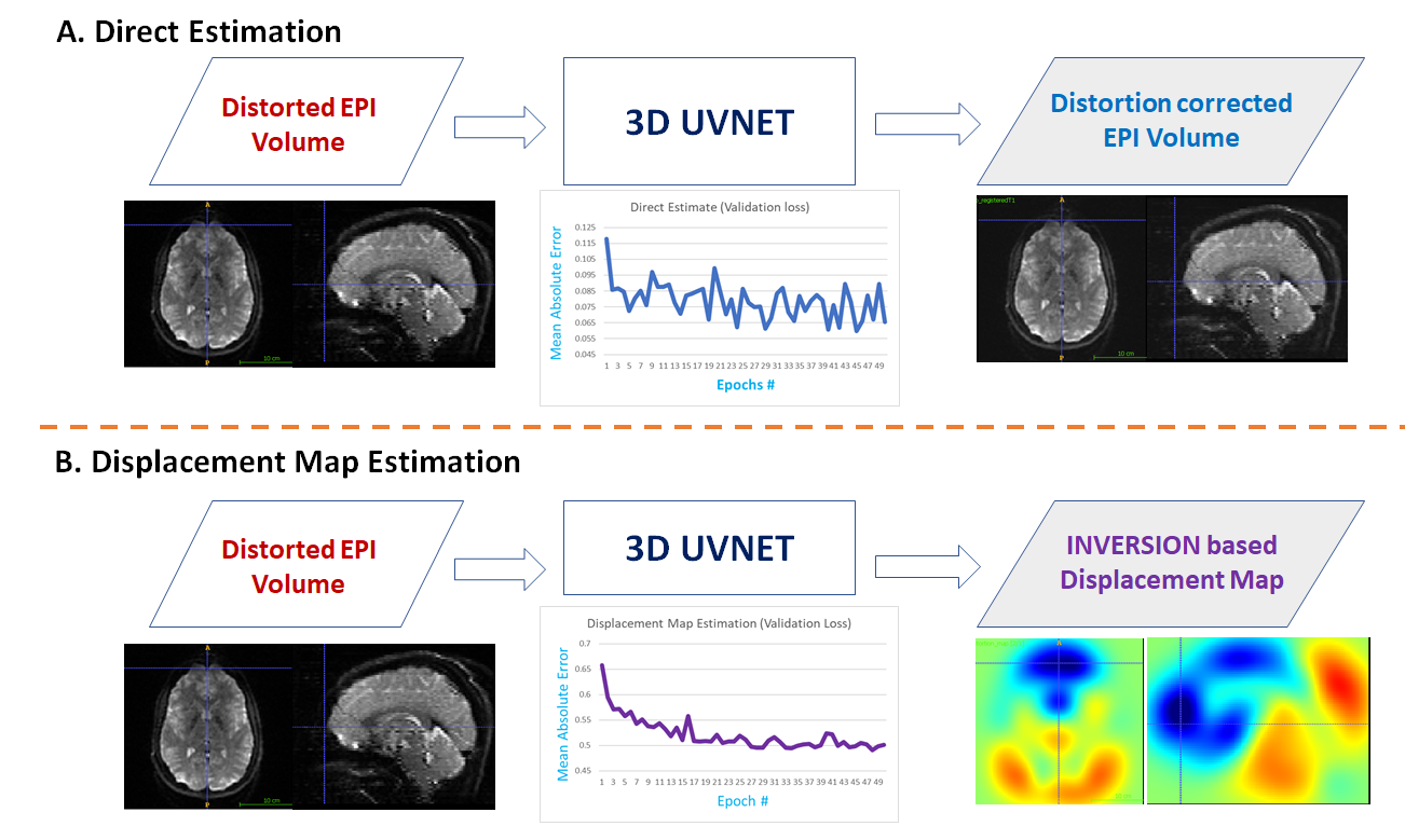
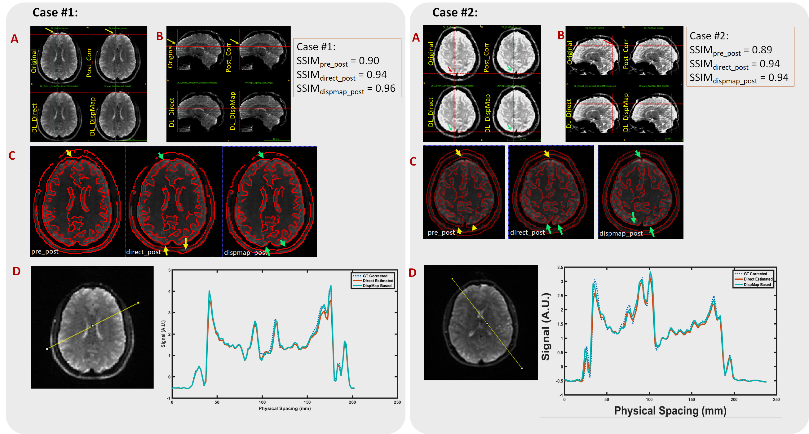
Ground truth displacement map: We use distortion map computed by using an implementation of INVERSION [1] as ground-truth (GT). INVERSION uses a T1-w anatomical image of subject as undistorted reference for correcting T2w EPI image [1]. Computed displacement map is used to generate reference corrected EPI images (Post_Corr). DL Experiment is described in Fig.1.
a. Direct Distortion Correction: A deep learning framework to directly estimate the distortion corrected EPI- volume; with distorted EPI-volume as input. This is like DL_Direct [2].
b. Displacement map estimation and distortion correction (DL_DispMap): Same deep learning framework is used to estimate displacement map from INVERSION method [1]. Input to DL- network is a EPI distorted volume. DL predicted displacement map is used to un-warp distorted volume by applying appropriate transform.
c. DL Architecture and training: We adapted a 3D UV-Net [3] to mitigate aliasing and smoothing artifacts due to down-sampling [5-8] for both experiments (Fig 1, a and b) and implemented in Keras [4]. Overall, 818 exams were used for training and validation and 29 exams for testing. Model with least validation loss was retained as the best model.
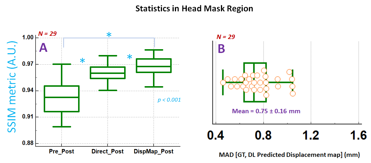
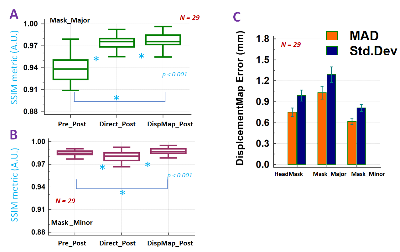
Accuracy Assessment: Assessment was done visually and quantified with Structural Similarity index (SSIM) metric [2] and displacement map error metrics. Head mask was obtained and further segmented into two regions: Mask_Major where GT displacement map > 1.875 mm and Mask_Minor (GT dispmap <= 1.875 mm). We choose the Post_Corr volume as reference and computed SSIM metrics with original EPI distorted volume (SSIMpre_post), and DL-based distortion corrected volumes from experiment a. (SSIMdirect_post) and b. (SSIMdispmap_post). Metrics are computed in all three masks.
Interpretation: SSIM ≈ 1 and displacement map error < 1.875 mm is considered a close match with Post_Corr. It is also expected that most of the changes should occur in Mask_Major region and minimal correction in Mask_Minor region.
Results and Discussion:
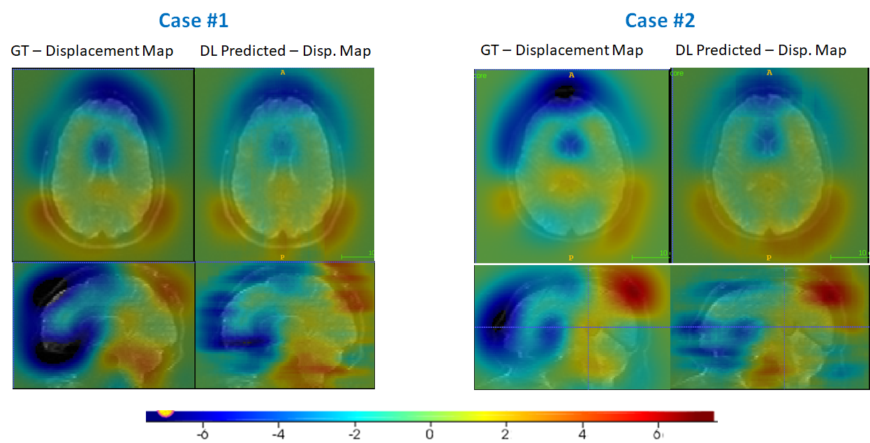
Smoother loss converge for DL_DispMap compared to direct estimation suggests better training performance for map estimation (Fig 1);. Overall, while both methods can correct distortion (Fig.2A, B); DL_Direct volumes tend to lose textural details as compared to volumes obtained from DL_DispMap method (Figs. 2D, profile plot). The DL predicted displacement maps show good overlap in focal spots (i.e. regions in Mask_Major) (Fig. 3). SSIM metric value is highest for DL_DispMap based correction (both in head mask as well as segmented regions) since it doesn't suffer from downsampling aliasing artifacts in image space as is the case with DL_Direct method (Fig.4A, Fig. 5A,B). For displacement map predicted by DL, we notice that MAD error (0.98 mm) is less than 1.875 mm for all the mask regions (Figs. 4B, 5C); indicating good accuracy. Overall, displacement map-based methodology allows for reasonable and consistent correction of EPI related distortion in brain MRI data. The method can potentially be also applied for fMRI studies where signal fidelity needs to be preserved.
One of the shortcoming of the method is that DL model is applicable only for an anatomy position/orientation and system configuration and might need to be re-retrained if either is changed.
Conclusion:
We establish that using displacement map-based method is better suitable to correct EPI distortion than direct correction approach. Corrected images demonstrate good match with gold standard INVERSION method (SSIM ≈ 0.97)Acknowledgements
No acknowledgement found.References
[1]. Bhushan C, Haldar J.P, Choi S, et.al.; Neuroimage. 2015 ; 115: 269–280.
[2]. P. Liao et al., Computers in Biology and Medicine 100 (2018): 230–238
[3]. Milletari, Fausto & Navab, Nassir & Ahmadi, Seyed-Ahmad. (2016). V-Net: Fully Convolutional Neural Networks for Volumetric Medical Image Segmentation. 565-571. 10.1109/3DV.2016.79.
[4]. https://keras.io
[5]. Tobias Springenberg et.al., https://arxiv.org/pdf/1412.6806.pdf
[6]. T. Tong, G. Li, X. Liu and Q. Gao, 2017 ICCV, Venice, 2017, pp. 4809-4817.
[7]. Xiao-Jiao Mao et.al; 2016. Proceedings NIPS'16, USA, 2810-2818.
[8]. B. Lim, S. Son, H. Kim, S. Nah and K. M. Lee, 2017 IEEE CVPRW, Honolulu, HI, 2017, pp. 1132-1140.
Figures