3402
Learned Gibbs Removal in Partial Fourier Acquisitions for Diffusion MRI1Radiology, NYU School of Medicine, New York, NY, United States
Synopsis
Despite significant advances in both denoising and Gibbs artifact removal, in acquisitions such as partial Fourier encoding, noise and Gibbs ringing continue to be an issue. Here we demonstrate that a machine learning approach can extend Gibbs ringing and noise removal to partial Fourier image acquisitions and show results on estimates of diffusion parameters on phantom and brain imaging data.
Introduction
Over the last few years, convolutional neural networks (CNNs) have become one of the most powerful image processing tools in MRI. One potential application of CNNs in MRI is Gibbs artifact removal. Gibbs ringing can induce bias in quantitative methods such as in diffusion parameter mapping.1 Sophisticated correction methods have been developed, but these methods may not extend to nonstandard acquisitions such as partial Fourier (PF).2 CNN-based Gibbs removal has shown promise relative to these methods;3,4 however, relatively few studies have examined whether CNN-based Gibbs removal actually leads to an improvement in the image-derived parameters of interest. Here, we attempt to address this gap by investigating the effects of CNN-based Gibbs removal in two experiments. The first is a highly-controlled water phantom experiment, while the second involves in vivo brain mapping of diffusion parameters. In the first case, the true kurtosis value of 0 is known, so we use this experiment as a validation of parameters derived from CNN DWIs. In the second experiment, we examine whether a neural network can accomplish the same task in vivo, using fully-sampled non-PF parameter maps as a reference.Methods
Neural Network Structure and Training: We used a 10-layer residual U-Net with modifications to improve resolution.5 Training was accomplished by using ImageNet, which has over 14 million images. Input training images were created by resizing the original ImageNet images to 256x256, cropping a 128x128 k-space block, adding noise, applying inverse FFT, and then taking the absolute value. Target images were created by spline-interpolating the original 256x256 images on to a 128x128 grid. In addition to this pipeline, data augmentation was accomplished by simulating random phase, random cropping, and varying SNR levels from 1 to 32. To simulate a partial Fourier acquisition, a mask was applied to the cropped k-space prior to the inverse FFT operation, the mask size depending on the PF factor (5/8ths in this case).
Comparison Methods: As a comparison, we used state-of-the-art (SoA) methods for denoising and deGibbsing. A subvoxel sinc shifting method was used correct for Gibbs artifacts.2 Since the neural network can perform both noise removal and Gibbs removal, prior to using subvoxel shifting, we applied a random matrix denoising routine (MPPCA).6,7
Validation Experiment: To validate the parameter maps from CNN preprocessing, we created a water phantom in which a central beaker of water was immersed in a larger water container. The wall of the beaker induced Gibbs artifacts. We measured diffusion weighted images with b-values of 500 s/mm2, 1000 s/mm2 and 1,500 s/mm2 along 66 isotropically distributed directions at a resolution of 1.89 mm by 1.89 mm by 1.50 mm. Since the kurtosis of water is 0, a ground truth is known for this experiment.
In Vivo Diffusion Experiment: The above sequence was repeated for in vivo diffusion measurements. In this case, Gibbs artifacts were present in mean diffusivity measurements, and we attempted removal with both the CNN method and random matrix denoising followed by subvoxel shifting.
Results
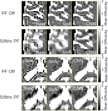
In our figures we show parameter maps from three classes: the raw diffusion-weighted images (Raw), the random matrix denoising with subvoxel shifting for deGibbsing (SoA - state-of-the-art), and the CNN-based method (CNN).
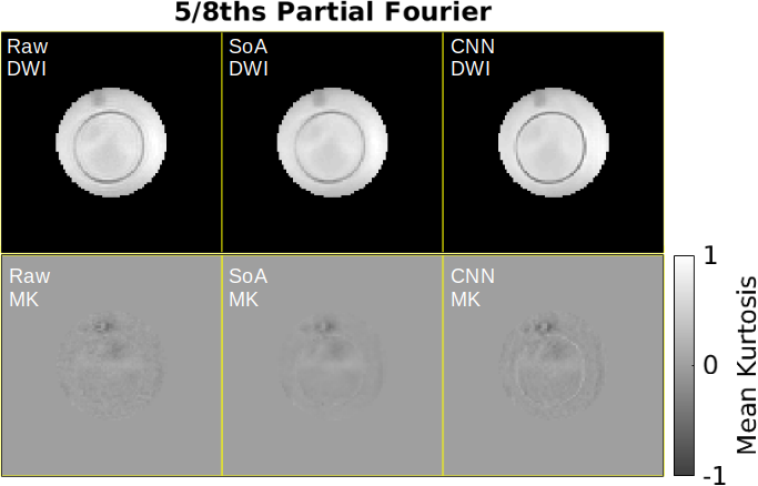
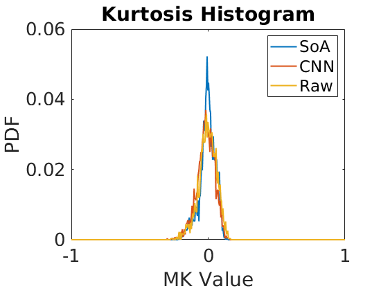
Figure 1 shows signal maps and mean kurtosis images from different methods of Gibbs removal. Denoising of the raw diffusion-weighted images also denoises the resultant MK maps. The histograms in Figure 2 show the skew of MK values that results from Gibbs artifacts and noisy data. Figures 1 and 2 indicate that all correction methods are able to estimate the true Kurtosis value of 0. Some mild residual blurring is present in the SoA DWI image in Figure 1 as it doesn't completely remove the partial Fourier effects.
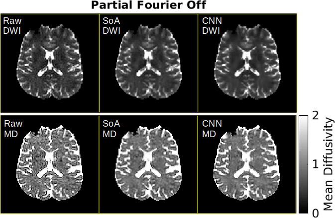
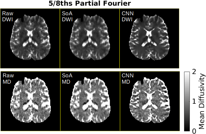
In vivo data from acquisitions without partial Fourier (Figure 3) and with partial Fourier (Figure 4) show Gibbs artifacts around the lateral ventricles in mean diffusivity measurements. The SoA method struggles to remove these artifacts in the presence of partial Fourier, but the CNN method accomplishes greater Gibbs reduction around the lateral ventricles as demonstrated in the detail images in Figure 5. In addition, the CNN achieves some resolution enhancement of features lost due to the partial Fourier acquisition (also in Figure 5).
Conclusions
CNN-based Gibbs removal methods can successfully both remove Gibbs artifacts and enhance resolution in partial Fourier acquisitions. Since the CNN only operates on individual, 2D images, it can be applied to data-starved protocols that are limited in the number of available diffusion directions or receive coils. The CNN-based approach is flexible and can be applied to a variety of non-standard acquisitions in which explicit modeling of image artifacts is difficult.
Acknowledgements
We would like to thank Greg Lemberskiy for aiding in the collection of the in vivo data. We would like to thank NIH R01 EB024532 and NIH P41 EB017183 for funding for this projectReferences
1. Veraart, J., Fieremans, E., Jelescu, I. O., Knoll, F., & Novikov, D. S. (2016). Gibbs ringing in diffusion MRI. Magnetic resonance in medicine, 76(1), 301-314.
2. Kellner, E., Dhital, B., Kiselev, V. G., & Reisert, M. (2016). Gibbs‐ringing artifact removal based on local subvoxel‐shifts. Magnetic resonance in medicine, 76(5), 1574-1581.
3. Wang, Y., Song, Y., Xie, H., Li, W., Hu, B., & Yang, G., Reduction of Gibbs artifacts in magnetic resonance imaging based on Convolutional Neural Network, CISP-BMEI 2017
4. Zhang, Q., Ruan, G., Yang, W., Zhao, Wu, E., & Feng, Y., Gibbs-Ringing Artifact Reduction in MRI via Machine Learning Using Convolutional Neural Network, ISMRM 2018
5. Ye, J. C., Han, Y., & Cha, E. (2018). Deep convolutional framelets: A general deep learning framework for inverse problems. SIAM Journal on Imaging Sciences, 11(2), 991-1048.
6. Veraart, J., Fieremans, E., & Novikov, D. S. (2016). Diffusion MRI noise mapping using random matrix theory. Magnetic resonance in medicine, 76(5), 1582-1593.
7. Veraart, J., Novikov, D. S., Christiaens, D., Ades-Aron, B., Sijbers, J., & Fieremans, E. (2016). Denoising of diffusion MRI using random matrix theory. NeuroImage, 142, 394-406.
Figures