3398
Effects of Gradient Nonlinearities on Reproducibility and Accuracy of Diffusion MRI Metrics in the Brain.1Quantitative Medical Imaging/NIBIB, National Institutes of Health, Bethesda, MD, United States
Synopsis
Gradient nonlinearities in MRI cause spatially-varying b-values and diffusion gradient directions. In this work, we analyze whether these nonlinearities have a significant impact on data reproducibility and accuracy for brain studies. Our results indicate that not only FA and TR values have an increasing bias away from the isocenter of the magnet, but also differences in subject positioning and head orientation combined with nonlinearities have a significant effect on reproducibility. The effects were also observed in principal eigenvector directions computed with the tensor model.
Introduction
Gradient nonlinearities (GNL) in MRI systems lead to both spatial distortions in images1 and spatially-varying deviations of the b-values and diffusion gradient directions from those prescribed2-5. These deviations are typically ignored on clinical scanners, however, they have been shown to the affect accuracy of diffusion-derived metrics3. Moreover, GNL may affect reproducibility across scanning sessions even on the same scanner due to differences in subject positioning and head orientation. In this work, we analyze whether GNL have a significant impact on diffusion metrics reproducibility and accuracy for brain studies.Materials
Six subjects were scanned with the same protocol on five different days on a Philips Achieva 3T MRI system. Diffusion weighted images (DWIs) were acquired with typical parameters of clinical acquisitions (single-shot spin-echo, 10 volumes with low b-values, and 32 volumes with a b-value of 1100s/mm2, 32-channel coil, sense factor=2). This acquisition scheme was repeated for both AP and PA phase encoding directions, and a T2-weighted turbo spin-echo image was also acquired with 1.7mm isotropic resolution to enable robust EPI distortion correction.
Methods
1) Data processing with gradient nonlinearity correction: We computed GNL fields from measurements on a PVP solution6. On a separate set of experiments, it was shown the spherical harmonic coefficients obtained with this method are virtually identical to those of the manufacturer7, which however may not be accessible by the user. These coefficients were used to generate a spatially-varying B-matrix image in the native space of the b=0s/mm2 image. DWIs were subsequently corrected for motion and eddy-currents distortions and a new B-matrix image for each DWI was generated accounting for motion effects. EPI distortions were corrected using AP and PA encoded data8,9 and all DWIs were reoriented onto a structural image. For each subject, the same structural image was used as reorientation target ensuring alignment across sessions. B-matrix reorientation was subsequently performed for all B-matrix images.
2) Analysis: For each scan, diffusion tensors were estimated using nonlinear regression once with a spatially constant B-matrix (uncorrected) and once with the corresponding voxelwise B-matrices (corrected). Fractional anisotropy (FA), Trace (TR) (equivalent to 3 times the mean diffusivity) and the principal eigenvector (e1) orientation dispersion (PEOD) metric10 were used for statistical testing. A voxelwise t-test was performed using the five scans of each subject to test $$$H_0:\mu^{{corrected}}=\mu^{{uncorrected}}$$$. All statistical maps were warped into a representative atlas space computed using DR-TAMAS11 to display the effects that could be expected in a population analysis.
Results
Figure 1 shows maps for the uncorrected and corrected diffusion metrics in a representative subject. As expected effects of nonlinearities were more pronounced in regions farther from the isocenter of the magnet. For TR errors were up to 8% and for FA up to 3%.
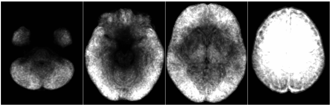
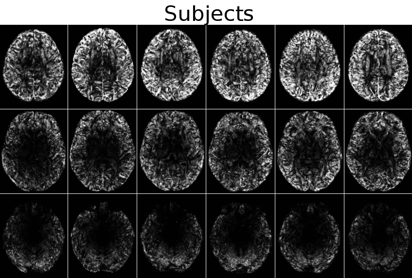
Figure 2 shows (1-p_values) maps obtained from an unpaired t-test on corrected vs. non corrected TR values for each subject. These maps essentially show in each voxel the likelihood of finding spurious statistically significant differences in longitudinal scans because of gradient nonlinearities. Not surprisingly, the likelihood of finding these spurious differences increases farther from the magnet isocenter. Interestingly, there is a halo-like band of lower probability in different locations for each subject. These bands reflect the effect of variability in repositioning subjects in repeated scans and originate from a relatively large variance in voxelwise B-matrices. Figure 3 displays the average of all maps warped into population atlas space. Averaging across a population reduces the effects of single subject repositioning; therefore the population averaged maps more closely resemble the actual nonlinearity fields and the low probability bands are no longer detectable.
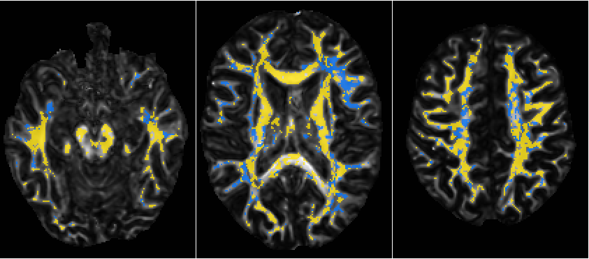
Figure 4 displays the maps for FA t-tests. Bands of varying probability are not observable for FA. One should not expect the same pattern for both TR and FA, because FA is also affected by the off-diagonal elements of B-matrices and not only its trace. Figure 5 displays the regions where e1 was more coherent among scans in orange and less coherent in blue after correction. Coherency is generally improved at different levels but this improvement becomes apparent near brain periphery.
Conclusions & Discussions
Our results indicated that gradient nonlinearities combined with differences in subject positioning can have a significant impact not only on the accuracy of diffusion MRI metrics, but also on their reproducibility. The statistical manifestations of these effects have a non-obvious spatial pattern, particularly for longitudinal scans. In general effects are more pronounced at the periphery of the brain, impairing one's ability to asses subtle cortical and sub-cortical diffusivity changes, as it is needed for example in TBI research. Gradient nonlinearities were also shown to alter the reproducibility of the directional information in sub-cortical white matter regions with potential implications for tractography and connectivity analyses.
Acknowledgements
Support included funding from the intramural program of the National Institute of Biomedical Imaging and Bioengineering in NIH.References
1. Glover GH and Pelc NJ. "Method for correcting image distortion due to gradient nonuniformity". US Patent # 4,591,789, May 27, 1986.
2. Dariya I. Malyarenko, Brian D. Ross, and Thomas L. Chenevert, "Analysis and correction of gradient nonlinearity bias in apparent diffusion coefficient measurements". Magn Reson Med. 2014;71(3):1312-23.
3. Malyarenko D, Galbán CJ, Londy FJ, Meyer CR, Johnson TD, Rehemtulla A, Ross BD, Chenevert TL, "Multi-system repeatability and reproducibility of apparent diffusion coefficient measurement using an ice-water phantom", J Magn Reson Imaging. 2013 May;37(5):1238-46.
4. Tan ET, Marinelli L, Slavens ZW, King KF, Hardy CJ., "Improved correction for gradient nonlinearity effects in diffusion-weighted imaging", J Magn Reson Imaging. 2013 Aug;38(2):448-53.
5. Bammer R1, Markl M, Barnett A, Acar B, Alley MT, Pelc NJ, Glover GH, Moseley ME. "Analysis and generalized correction of the effect of spatial gradient field distortions in diffusion-weighted imaging". Magn Reson Med. 2003 Sep;50(3):560-9.
6. Pierpaoli C, Sarlls J, Nevo U, Basser PJ, Horkay F. Polyvinylpyrrolidone (PVP) water solutions as isotropic phantoms for diffusion MRI studies. Honolulu, Hawai'i: 2009. p. 1414.
7. ISMRM 2019, abstract #4044 (submitted).
8. Pierpaoli C, Walker L, Irfanoglu MO, Barnett AS, Chang LC, Koay CG, et al. TORTOISE: An integrated software package for processing of diffusion MRI data. In: Proceedings of International Society of Magnetic Resonance in Medicine; 2010.p. 1597.
9. Irfanoglu MO, Modi P, Nayak A, Hutchinson EB, Sarlls J, Pierpaoli C. DR-BUDDI: (Diffeomorphic Registration for Blip-Upblip-Down Diffusion Imaging) method for correcting echo planar imaging distortions. Neuroimage 2015;106:284–289.
10. Basser PJ, Pajevic S. Statistical artifacts in diffusion tensor MRI (DTMRI) caused by background noise. Magnetic Resonance in Medicine. 2000;44:41–50.
11. Irfanoglu MO, Nayak A, Jenkins J, et al. DR-TAMAS: Diffeomorphic Registration for Tensor Accurate Alignment of Anatomical Structures. Neuroimage. 2016;132:439-454.
Figures
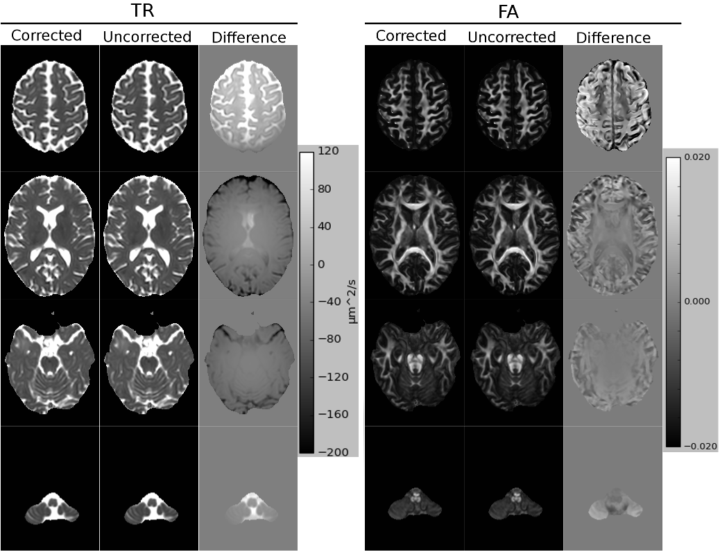
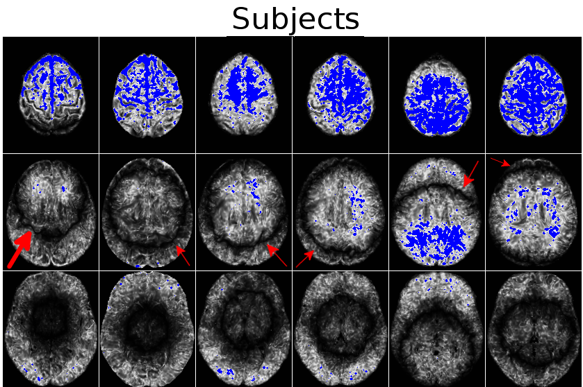
Figure 2. Maps obtained from t-tests between the TR values of corrected and uncorrected data for all subjects, at three slice levels. The underlays are the (1-p_value) significance maps and the overlayed blue voxels are statistically significant voxels at $$$\alpha=0.05$$$. Statistical significance increases again further from the center. However, bands of lower significance exist (red arrows) for all subjects.