3382
Altered structural connectivity in the auditory-related pathway in patients with idiopathic sudden sensorineural hearing loss by diffusion spectrum imaging1Department of Radiology, Beijing Chao-Yang Hospital, Capital Medical University, Beijing, China, 2MR Scientific Marketing, Siemens Healthcare, Beijing, China, 3Department of Hyperbaric Oxygen, Beijing Chao-Yang Hospital, Capital Medical University, Beijing, China
Synopsis
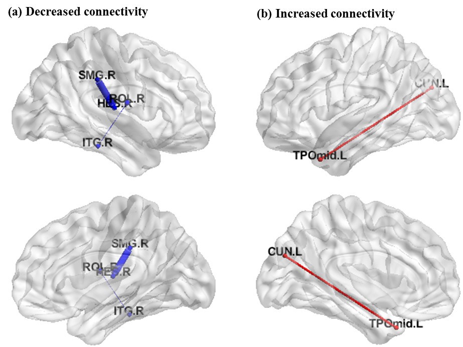
Sensorineural hearing loss is increasingly recognized as the result of alterations in the auditory-related network. The present study aimed to further explore the whole brain abnormalities of neural network connections in idiopathic sudden sensorineural hearing loss (ISSNHL) through diffusion spectrum imaging. It was found left-sided ISSNHL exhibited decreased connectivity between the contralateral inferior temporal gyrus and rolandic operculum, and the contralateral heschl gyrus and superior marginal gyrus; while increased connectivity was detected between the ipsilateral temporal pole and cuneus, which suggests that DSI could help investigate the structural correlates of these imaging abnormalities in this disease.
Introduction
Sensorineural hearing loss is a common emergency in the department of otolaryngology. It is increasingly recognized as the result of an altered brain network,[1,2]. both on the structural and functional levels. The characterization of these widespread brain alterations is crucial for our understanding of the clinical manifestation as well as for the treatment. Tractography based on Diffusion Tensor Imaging allows non-invasive mapping of white matter tracts in vivo. Recently, diffusion spectrum imaging (DSI),[3] based on an increased number of diffusion directions and intensities, has improved the sensitivity of tractography, notably with respect to the problem of fiber crossing and recent developments allow acquisition times compatible with clinical application. This study aimed to further explore the abnormalities of neural network connections in idiopathic sudden sensorineural hearing loss (ISSNHL) patients through generalized DSI.METHODS
30 patients with ISSNHL (right 14, left 16) were enrolled as the experimental group and 24 matched healthy volunteers were enrolled as the control group. These participants were performed MRI scans at 3.0T (MAGNETOM Prisma, Siemens, Erlangen, Germany) with a 64 channels head coil within two weeks after visiting outpatient firstly. The protocols included the DSI, T1-weighted magnetization-prepared rapid gradient echo (T1-Mprage) and 3D-FIASTA sequence in order to exclude inner ear malformation and other inner ear lesions. Echo-planar Imaging (EPI) q-space imaging were collected with the following parameters: TR = 7200 ms, TE = 79 ms, flip angle = 8°, field-of-view 220 × 220 mm2, bmax 3000 s/mm2, direction 257 and voxel size 2.2×2.2×2.2 mm3. T1-Mprage sagittal images were collected with the following parameters: TR = 2300 ms, TE = 2.27 ms, flip angle = 8°, field-of-view 256 × 256 mm2, 192 slices, and voxel size 1×1×1 mm3. This study followed the framework of diffusion spectrum imaging (DSI) in which diffusion weighted images were acquired with diffusion gradients of different b values corresponding to the grid points filled within a sphere in the 3D diffusion-encoding space (q-space). DSI data were preprocessed by DSI studio (http://dsi-studio.labsolver.org/) and whole brain structural connectivity matrices were built based on quantitative anisotropy (QA) between 90 brain regions of interest (ROI) from the Automated Anatomical Labeling (AAL) template using GRETNA software.[4] Two-sample t-test was performed to determine which connectivity was significantly altered between in patients with ISSNHL in comparison to healthy controls. Results were thresholded at p < 0.05 FWE-corrected.RESULTS
In comparison with healthy controls, the left-sided ISSNHL patients exhibited significantly reduced connections between the contralateral rolandic operculum and inferior temporal gyrus (T = -3.58), and the contralateral supramarginal and Heschl's gyrus (T = -3.61); while the connection between the ipsilateral temporal pole and cuneus were significantly increased (T = 3.99) in ISSNHL (see Figure 1). The similar pattern could be found in patients with right-sided ISSNHL that the reduced connection was identified between the contralateral parahippocampal gyrus and angular (T = -3.70).DISCUSSION
Our study is the first application of tractography computed from DSI data, a high angular resolution diffusion technique, for investigating whole-brain structural connectivity changes in patients with ISSNHL. We found connectivity alterations in contralateral decrease and ipsilateral increase in ISSNHL patients, notably in the temporal lobe which is considered as auditory nerve center.[5] We found both an intra-temporal and extratemporal alteration which can be explained as a structural connectivity based on the anisotropy of the diffusion.CONCLUSION
Our study provides further structural evidence of injury on the opposite side of the auditory nerve center, compensation of the hearing loss side auditory nerve center and DSI-based imaging could help investigate the structural correlate of these imaging abnormalities.Acknowledgements
No acknowledgement found.References
1. Chang Y, Lee SH, Lee YJ, et al. Auditory neural pathway evaluation on sensorineural hearing loss using diffusion tensor imaging. Neuroreport 2004; 15(11): 1699-1703.
2. Sang H L, Chang Y, Ji E L, et al. The values of diffusion tensor imaging and functional MRI in evaluating profound sensorineural hearing loss. Cochlear Implants International 2010; 5(S1):149-152.
3. Wedeen V J, Wang R P, Schmahmann J D, et al. Diffusion spectrum magnetic resonance imaging (DSI) tractography of crossing fibers. Neuroimage 2008; 41(4):1267-1277.
4. Wang J, Wang X, Xia M, et al. GRETNA: a graph theoretical network analysis toolbox for imaging connections. Front Hum Neurosci 2015; 9:386.
5. Deepak N. Pandya, Douglas L. Rosene, Andrew M. Doolittle. Corticothalamic connections of auditory-related areas of the temporal lobe in the rhesus monkey. The Journal of Comparative Neurology 2010; 345(3):447.
Figures