3372
TractEM: A fast protocol for Whole Brain Tractography1Computer Science, Vanderbilt University, Nashville, TN, United States, 2Vanderbilt University Institue of Imaging Science, Nashville, TN, United States, 3Biomedical Engineering, Vanderbilt University, Nashville, TN, United States, 4Electrical Engineering, Vanderbilt University, Nashville, TN, United States, 5Laboratory of Behavioral Neuroscience, National Institue of Aging, National Institutes of Health, Baltimore, MD, United States, 6Radiology and Radiological Sciences, Vanderbilt University, Nashville, TN, United States
Synopsis
We introduce TractEM, a tractography-based whole-brain labeling protocol informed by the Eve Labeling [1] procedures from the single-subject Johns Hopkins white matter atlas [1, 2]. This project proposes to create a resource of manually labelled white matter atlases that is driven by state-of-the-art diffusion tractography, and can be manually created in less than 6 hours. We defined and tested the TractEM protocol on 61 tracts for 20 subjects, with multiple raters per subject, and show moderate to high reproducibility for most labels. TractEM should be a useful resource for generating target templates for automated labeling methods.
Introduction
Reproducible identification of white matter tracts across subjects is essential for the study of the structural connectivity of the human brain. Typically, labelling single-subject white matter tracts is done by propagating an atlas (or multiple atlases) to unlabeled data [3]; however, most digital atlases treat white matter as essentially homogenous (as white matter exhibits isointense signal on structural MRI). The state-of-the-art white matter atlas is the single-subject Johns Hopkins atlas [1], labeled using the Eve protocol using both diffusion tensor data and a T1-structural image of the brain. However, only a single subject was labelled, and manual delineation (at the time) was extremely time consuming. Additional independent subjects labeled with equivalent methodologies, and the tools and protocols to do so, would be useful resources for the neuroimaging community. This project aims to provide sets of manually defined labels for regional white matter definitions by developing a protocol informed by the Eve labeling scheme so that (1) all pathways can be manually traced in less than 6 hours, (2) using regions driven by current state-of-the art tractography, (3) with a methodology that can be applied generally to a large number of subjects.Methods
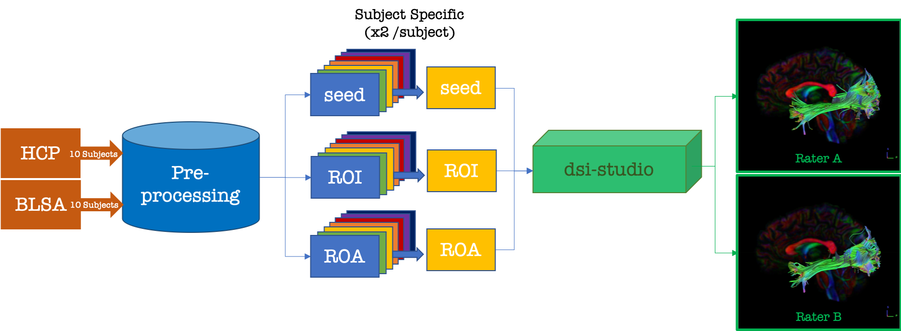
The TractEM protocol was developed and tested on 10 subjects from the HCP project [4] (age range 26-36) and 10 subjects from the BLSA project [5] (age range 57-77). Figure 1 shows the general processing pipeline which includes standard diffusion pre-processing, affine registration to JHU-Talairach space [1, 6], voxel-wise reconstruction using Q-ball (or generalized Q-ball for multi-shell HCP data) imaging [7], and tract generation using DSI studio software and unique combinations of seed-regions, ROI-regions, and not-regions (ROA’s). Full processing code can be found at https://github.com/MASILab/tractem/preprocessing, and detailed tracking protocols at https://my.vanderbilt.edu/tractem/protocol/.Results
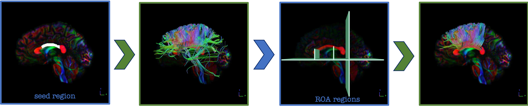
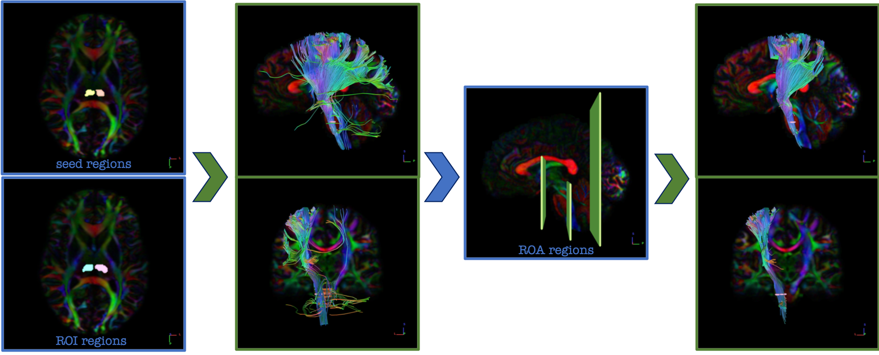
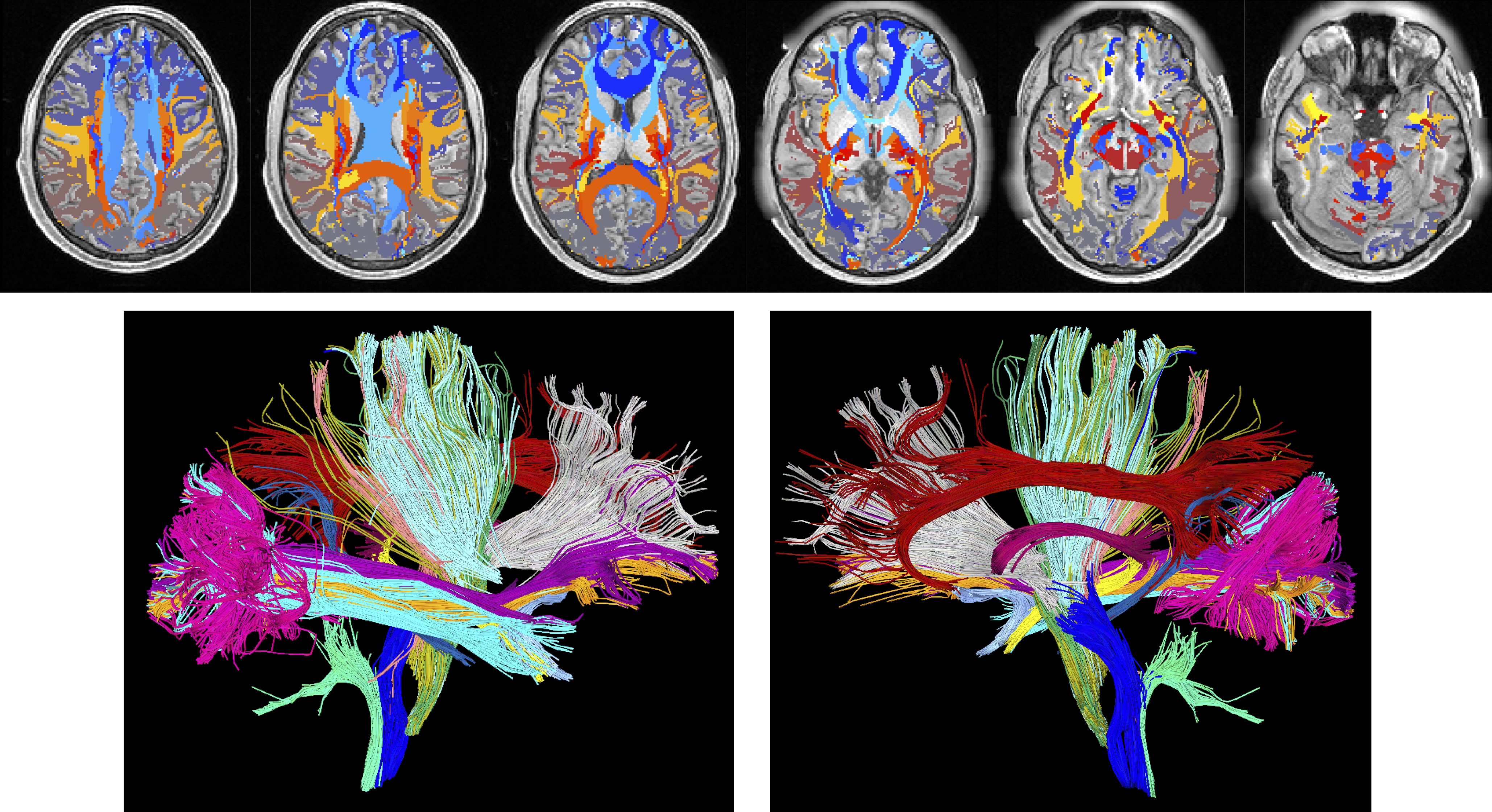
61 white matter pathways were generated, twice per subject, with TractEM protocol documented as PDFs for each pathway (https://my.vanderbilt.edu/tractem/protocol/). Example protocols and tractography results are shown for the genu of the corpus callosum (Figure 2) and the corticospinal tract (Figure 3), which display tract-specific seed-regions, ROI-regions, and ROA-regions. A 3D visualization of a number of white matter pathways and labels is shown in Figure 4.
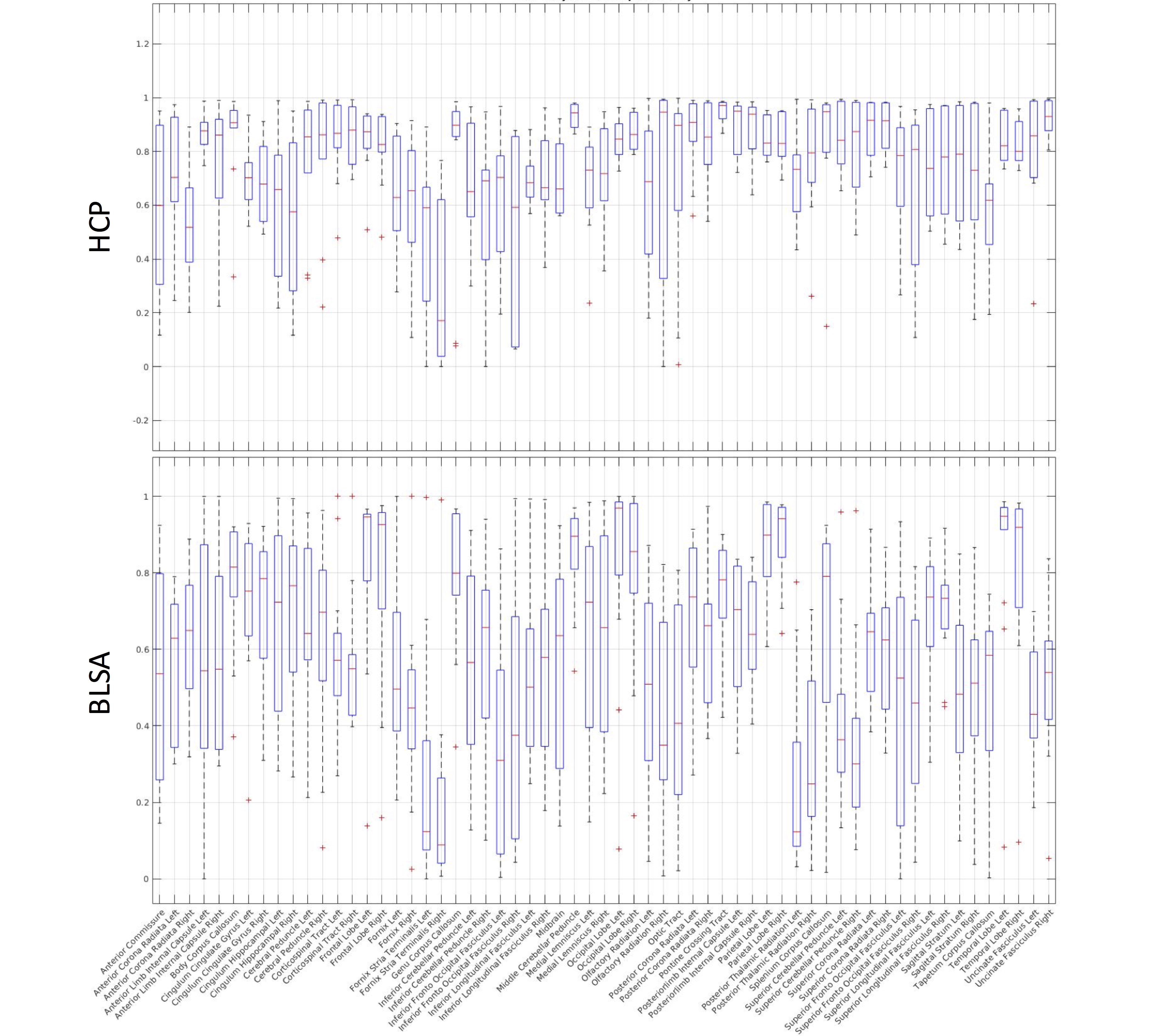
Reproducibility is assessed using the Dice overlap coefficient. Figure 5 shows the reproducibility across raters for each white matter label, for 10 subjects from each HCP (top) and BLSA (bottom) datasets. In general, the high-quality acquisition of the HCP datasets resulted in greater reproducibility than BLSA. Both datasets show similar trends, with larger pathways being generally more reproducible than smaller ones.
Discussion
The creation of manually labeled white matter atlases could serve as valuable neuroscience resources for use as target templates and for automated labeling methods. Currently, very few methods segment white matter regions, and those that do typically have few (or single) subjects, or limited white matter labels [1, 8, 9]. Here, we introduce a protocol for tractography-based whole-brain labeling that follows state-of-the-art white matter definitions, and can be performed for all 61 tracts in a matter of hours. Similar white matter segmentations using automated tractography methods are gaining popularity with ROI-based and clustering-based segmentation methods [10-12].
The tracking protocol is freely available (https://my.vanderbilt.edu/tractem/protocol/), documented in electronic reports, and version controlled. We welcome feedback and edits on particular tract protocols as well as high level processing. Future work will include labeling more datasets, investigating reasons for low reproducibility, and exploring automated segmentations using multi-atlas, machine learning, and tractography-guided methodologies.
Acknowledgements
This work was supported by the National Institutes of Health under award numbers R01EB017230, and T32EB001628, and in part by ViSE/VICTR VR3029 and the National Center for Research Resources, Grant UL1 RR024975-01. This research was conducted with the support from Intramural Research Program, National Institute on Aging, NIH. The content is solely the responsibility of the authors and does not necessarily represent the official views of the NIH.References
1. Mori S, Oishi K, Jiang H, Jiang L, Li X, Akhter K, et al. Stereotaxic white matter atlas based on diffusion tensor imaging in an ICBM template. NeuroImage. 2008;40(2):570-82. doi: 10.1016/j.neuroimage.2007.12.035. PubMed PMID: 18255316; PubMed Central PMCID: PMCPMC2478641.
2. Oishi K, Faria A, Jiang H, Li X, Akhter K, Zhang J, et al. Atlas-based whole brain white matter analysis using large deformation diffeomorphic metric mapping: application to normal elderly and Alzheimer's disease participants. NeuroImage. 2009;46(2):486-99. PubMed PMID: 19385016; PubMed Central PMCID: PMCPMC2885858.
3. Asman AJ, Landman BA. Non-local statistical label fusion for multi-atlas segmentation. Med Image Anal. 2013;17(2):194-208. doi: 10.1016/j.media.2012.10.002. PubMed PMID: 23265798; PubMed Central PMCID: PMCPMC3648421.
4. Van Essen DC, Ugurbil K, Auerbach E, Barch D, Behrens TE, Bucholz R, et al. The Human Connectome Project: a data acquisition perspective. NeuroImage. 2012;62(4):2222-31. doi: 10.1016/j.neuroimage.2012.02.018. PubMed PMID: 22366334; PubMed Central PMCID: PMCPMC3606888.
5. Ferrucci L. The Baltimore Longitudinal Study of Aging (BLSA): a 50-year-long journey and plans for the future. J Gerontol A Biol Sci Med Sci. 2008;63(12):1416-9. PubMed PMID: 19126858; PubMed Central PMCID: PMCPMC5004590.
6. Talairach J, Tournoux P. Co-planar stereotaxic atlas of the human brain : 3-dimensional proportional system : an approach to cerebral imaging. Stuttgart ; New York: Georg Thieme; 1988. 122 p. p.
7. Descoteaux M, Angelino E, Fitzgibbons S, Deriche R. Regularized, fast, and robust analytical Q-ball imaging. Magnetic resonance in medicine : official journal of the Society of Magnetic Resonance in Medicine / Society of Magnetic Resonance in Medicine. 2007;58(3):497-510. Epub 2007/09/01. doi: 10.1002/mrm.21277. PubMed PMID: 17763358.
8. Mori S, van Zijl P. Human white matter atlas. Am J Psychiatry. 2007;164(7):1005. doi: 10.1176/ajp.2007.164.7.1005. PubMed PMID: 17606649.
9. Oishi K, Zilles K, Amunts K, Faria A, Jiang H, Li X, et al. Human brain white matter atlas: identification and assignment of common anatomical structures in superficial white matter. NeuroImage. 2008;43(3):447-57. doi: 10.1016/j.neuroimage.2008.07.009. PubMed PMID: 18692144; PubMed Central PMCID: PMCPMC2586008.
10. Wasserthal J, Neher P, Maier-Hein KH. TractSeg - Fast and accurate white matter tract segmentation. NeuroImage. 2018;183:239-53. doi: 10.1016/j.neuroimage.2018.07.070. PubMed PMID: 30086412.
11. Wassermann D, Makris N, Rathi Y, Shenton M, Kikinis R, Kubicki M, et al. The white matter query language: a novel approach for describing human white matter anatomy. Brain structure & function. 2016;221(9):4705-21. doi: 10.1007/s00429-015-1179-4. PubMed PMID: 26754839; PubMed Central PMCID: PMCPMC4940319.
12. Yendiki A, Panneck P, Srinivasan P, Stevens A, Zollei L, Augustinack J, et al. Automated probabilistic reconstruction of white-matter pathways in health and disease using an atlas of the underlying anatomy. Front Neuroinform. 2011;5:23. doi: 10.3389/fninf.2011.00023. PubMed PMID: 22016733; PubMed Central PMCID: PMCPMC3193073.
Figures