3370
Using HCP data to improve diffusion tractography in routine-quality data: Application to the virtual dissection of the SLF system1Athinoula A. Martinos Center for Biomedical Imaging Massachusetts General Hospital and Harvard Medical School, Charlestown, MA, United States, 2Athinoula A. Martinos Center for Biomedical Imaging Massachusetts General Hospital and Harvard Medical School, SOMERVILLE, MA, United States
Synopsis
The development of scanners with ultra-high gradients, spearheaded by the Human Connectome Project, has led to dramatic improvements in the spatial, angular, and diffusion resolution that is feasible for in vivo diffusion MRI acquisitions. Here we show that global probabilistic tractography with anatomical priors can be trained on such data, which can only be acquired on a handful of Connectome scanners worldwide, and improve the accuracy of tractography in more widely available, routine-quality diffusion data. We apply this method to reconstruct the three subcomponents of the SLF and show its superior accuracy compared to a conventional multi-ROI approach.
Introduction
Diffusion tractography allows us to investigate white matter (WM) architecture in vivo and non-invasively. Several methods have been proposed for tractography to delineate WM bundles known from the anatomical literature. Approaches that involve manual definition of regions of interest (ROIs) in individual space1 are time-intensive and require extensive anatomical knowledge on the part of the operator, limiting reproducibility and applicability to large datasets. Atlas-based, multi-ROI approaches2,3 automate this process but rely on accurate alignment between each individual and an atlas. Importantly, all these methods perform local tractography on the individual data and apply a posteriori rules based on manual or atlas-based ROIs. If the resolution or SNR of the individual’s data is not sufficient to delineate the bundles of interest in their entirety, the a posteriori ROIs will not fix this, even if they are obtained from an atlas derived from very high-quality data. TRACULA (TRActs Constrained by UnderLined Anatomy)4 relies on a Bayesian framework for global tractography that incorporates prior probabilities on the anatomical neighborhood of WM pathways from a set of training subjects. The priors are included in the tract estimation itself, rather than as a posteriori deterministic rules. Here we show that this allows TRACULA to use high-quality training data to improve reconstruction in routine-quality data. We illustrate this in the superior longitudinal fasciculus (SLF) system. The SLF is a major fronto-parietal cortico-cortical association pathway with three subcomponents (SLFI, SLFII, SLFIII) organized in a medio-to-lateral and dorso-to-ventral fashion5. The human morphology of the most dorsal component (SLFI) remains controversial6,7, and its tractography-based reconstruction challenging. We use TRACULA, trained on high-b HCP data, to reconstruct the SLF system in lower-b data, and compare its performance to an automated multi-ROI approach.Methods
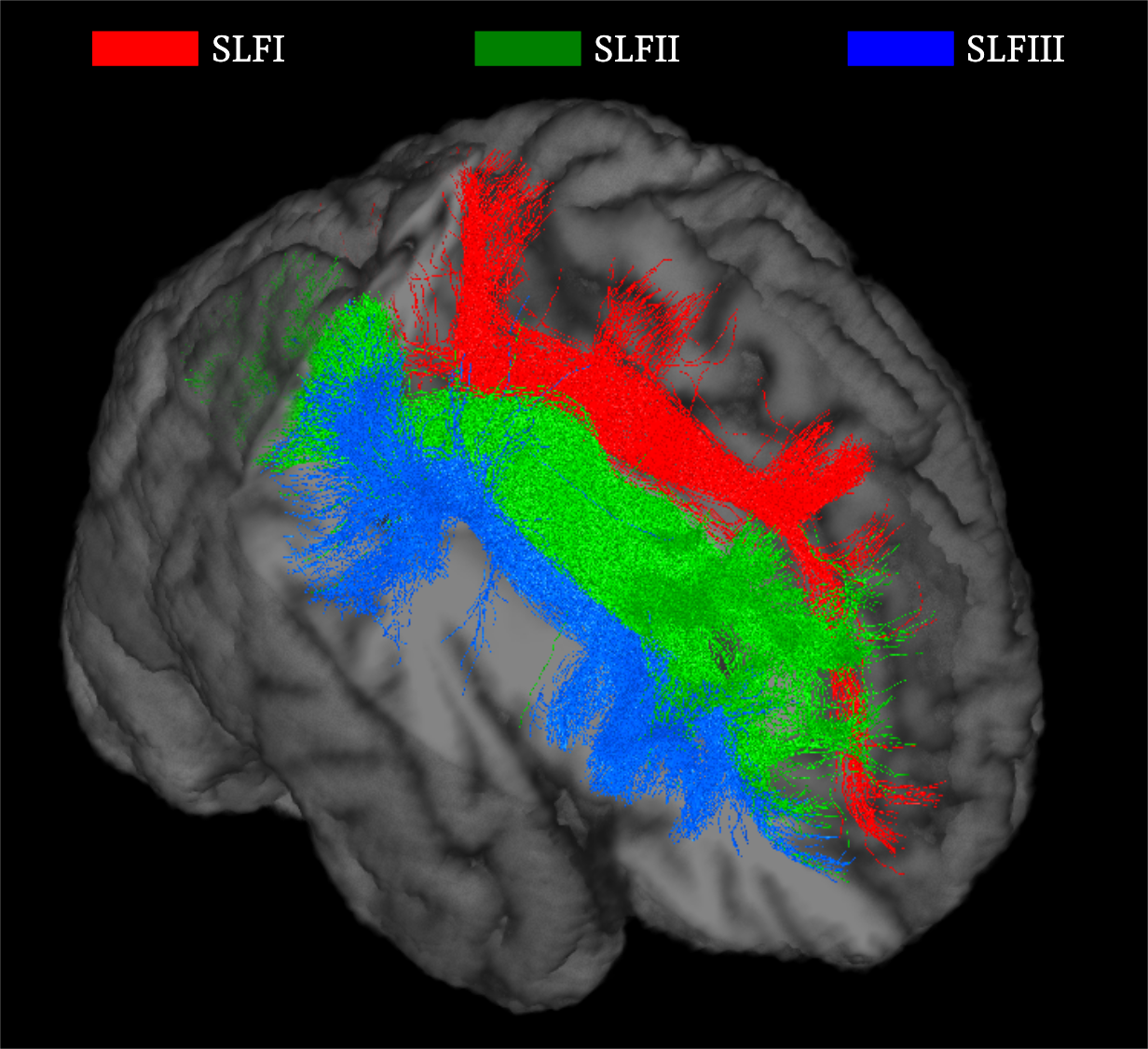
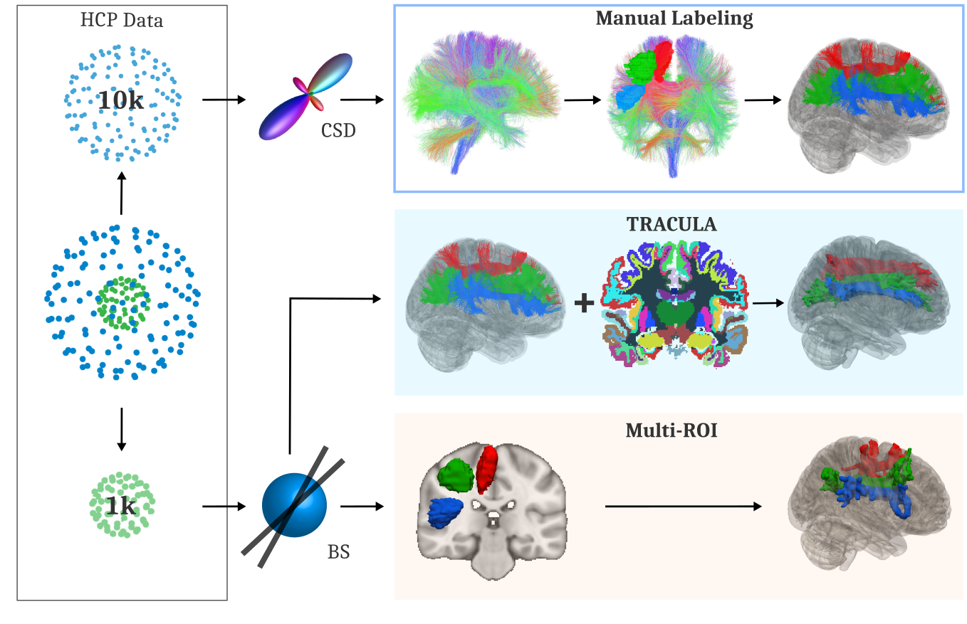
We use MGH-USC HCP data from 15 subjects8. Manual labeling: We perform whole-brain probabilistic tractography with constrained spherical deconvolution on the b=10000s/mm2 shell in DIPY9,10 (lmax=8, step-size: 0.5mm, angle-threshold: 30, 10 seeds/voxel). We dissect the SLF branches in Trackvis (Figure 1), following a protocol derived from the anatomical literature5,6,7 (author C.M.). SLFI: We place one ROI in the superior frontal gyrus and one encompassing the WM posterior to the posterior central gyrus and dorsal to the cingulate sulcus. SLFII: We place one ROI in the caudal part of the middle frontal gyrus and one in the WM of the inferior parietal lobe. SLFIII: We place one ROI in the inferior frontal gyrus and one in the supramarginal gyrus5,6,7. For all 3 bundles, we use mid-sagittal and temporal exclusion ROIs. TRACULA: For each subject, we use manually defined bundles from the other 14 subjects to obtain prior probabilities on the relative positions of the bundles with respect to the labels of an automated anatomical segmentation11,12. We apply TRACULA with these priors to reconstruct the bundles in the b=1000s/mm2 shell of the test subject. Multi-ROI method: We follow a common multi-ROI approach3. We register our manual ROIs to MNI space, average them, and map the average ROIs to the test subject. We use these as masks for local probabilistic tractography with the ball-and-stick model13, which is the model also used by TRACULA (Figure 2). Accuracy metrics: We compute the modified Hausdorff distance and Jaccard index between the pathways labeled manually in the high-b data and those extracted automatically, either by TRACULA or by the multi-ROI approach, in the low-b data of the same subject. We use thresholded (multi-ROI: the default 0.5%, TRACULA: 5%) and binarized distributions.Results
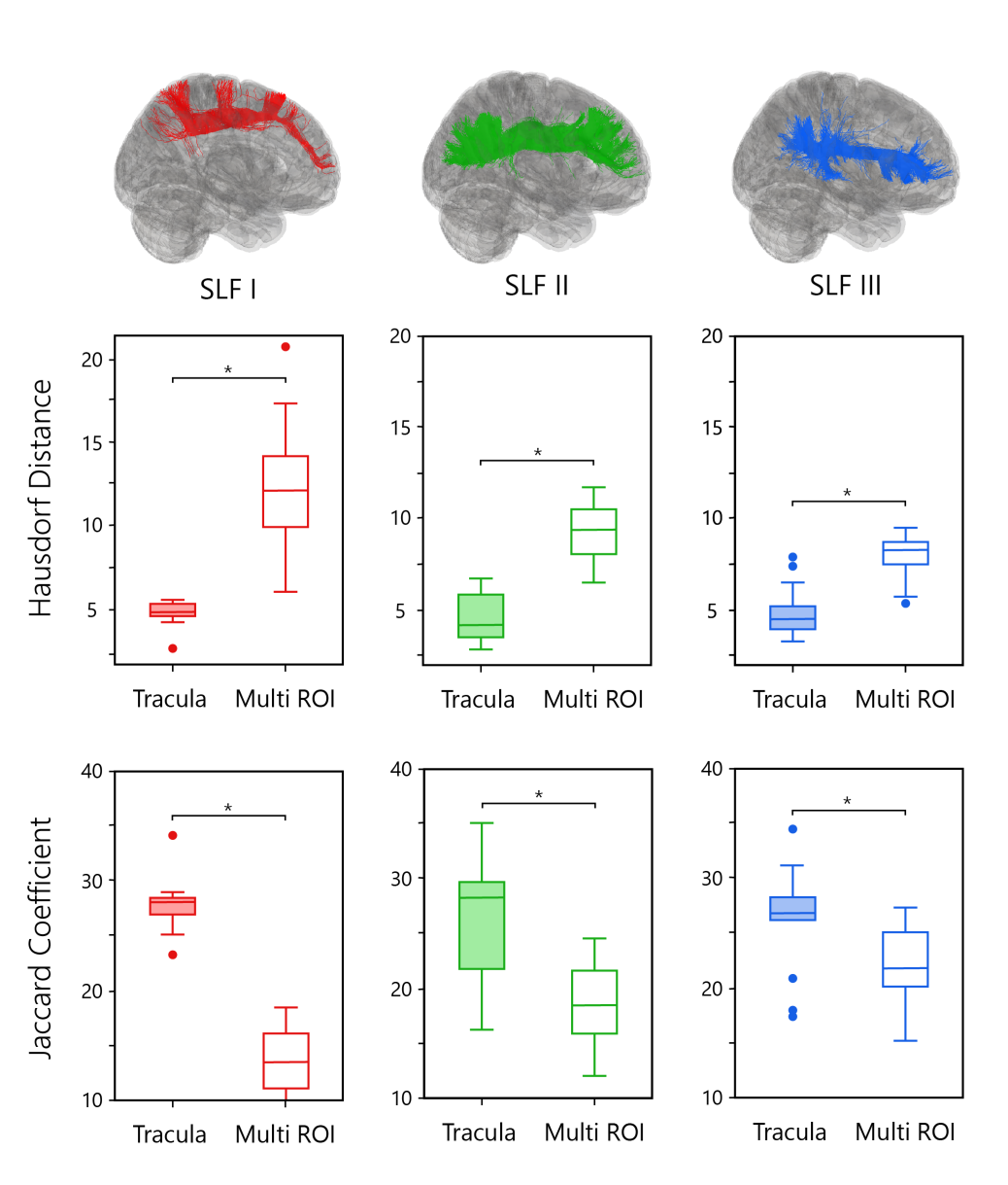
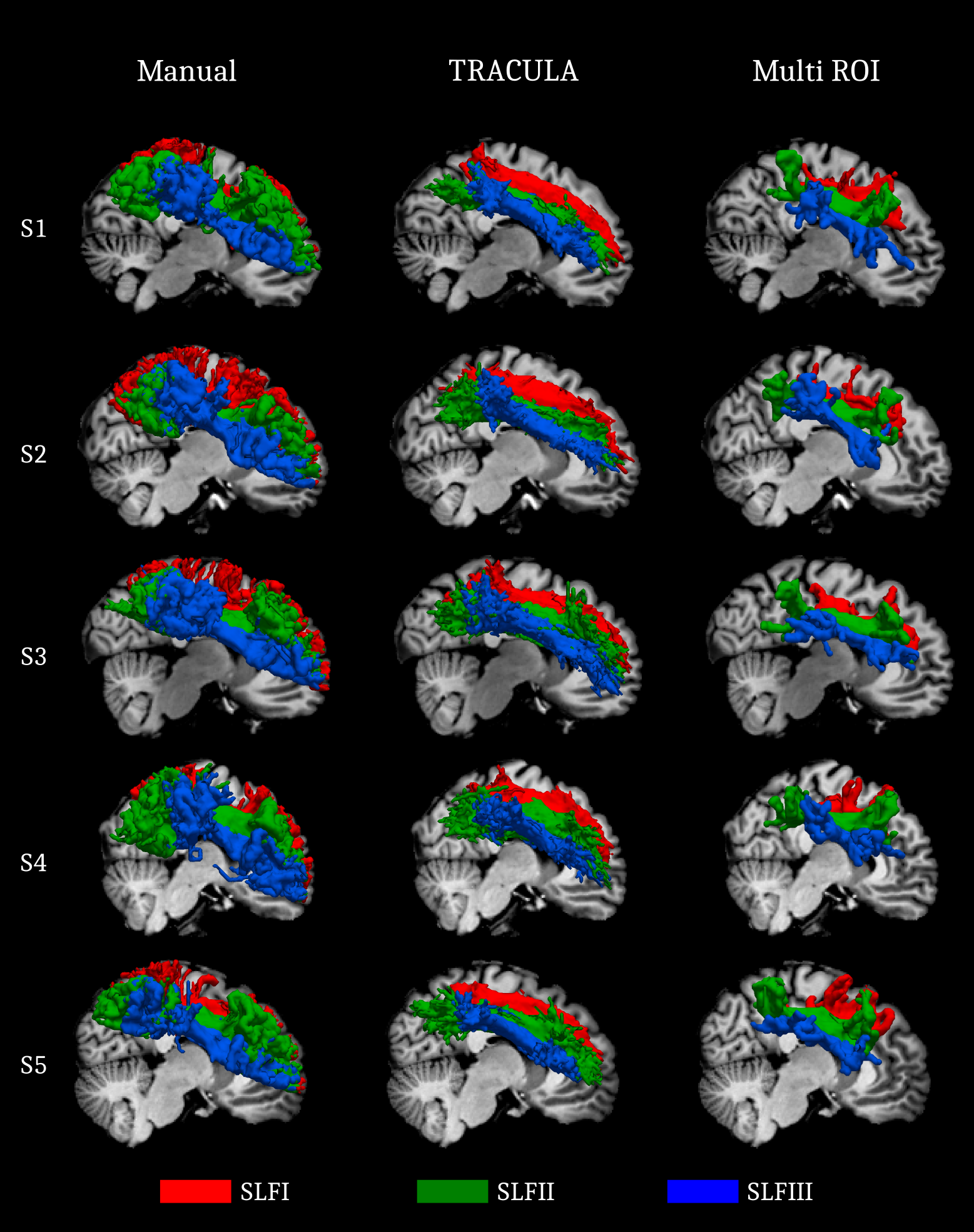
Both Hausdorff distance and Jaccard coefficient plots show significantly higher accuracy for TRACULA compared to the multi-ROI approach (Figure 3). Manual and automated reconstructions are shown in Figure 4. Lower accuracy for the multi-ROI approach is commonly due to not recovering the entire extent of the bundles. This is particularly true for the SLFI, which in most cases terminates near the precentral, central, and postcentral gyrus, instead of reaching the frontal cortex.Discussion
We have demonstrated that our global probabilistic tractography with anatomical priors can recover the three subcomponents of the SLF at low b-values by taking advantage of training data obtained at higher b-values. Importantly, it is able to do so with only a small number of training examples. Therefore we have illustrated its potential as a method for “data quality transfer” from limited-availability, high-quality data that can only be acquired on a handful of Connectome scanners worldwide, to more widely available, routine-quality data that can be acquired clinically. This allows the technological innovations of the HCP to benefit the wider community that does not have access to a Connectome scanner.Acknowledgements
This work was funded by NIH grant R01-EB021265.
Data were provided by the MGH-USC Human Connectome Project (U01-MH093765), funded by the NIH Blueprint for Neuroscience Research.
References
1. Catani M, Thiebaut de Schotten M. A diffusion tensor imaging tractography atlas for virtual in vivo dissections. Cortex. 2008 Sep;44(8):1105-32.
2. Yeatman JD, Dougherty RF, Myall NJ, Wandell BA, Feldman HM. Tract profiles of white matter properties: automating fiber-tract quantification. PLoS One. 2012;7(11):e49790.
3. De Groot, M., Vernooij, M.W., Klein, S., Ikram, M.A., Vos, F.M., Smith, S.M., Niessen, W.J., Andersson, J.L.R.. Improving alignment in Tract-based spatial statistics: Evaluation and optimization of image registration. NeuroImage. 2013;76, 400-411.
4. Yendiki A, Panneck P, Srinivasan P, et al. Automated probabilistic reconstruction of white-matter pathways in health and disease using an atlas of the underlying anatomy. Front Neuroinform. 2011;5:23.
5. Schmahmann JD, Pandya DN. Fiber Pathways of the Brain. Oxford University Press; 2006.
6. Hecht EE, Gutman DA, Bradley BA, Preuss TM, Stout D. Virtual dissection and comparative connectivity of the superior longitudinal fasciculus in chimpanzees and humans. Neuroimage. 2014;108:124-37.
7. Howells H, Thiebaut de Schotten M, Dell'Acqua F, et al. Frontoparietal Tracts Linked to Lateralized Hand Preference and Manual Specialization. Cereb Cortex. 2018;28(7):2482-2494.
8. Fan Q, Witzel T, Nummenmaa A, et al. MGH-USC Human Connectome Project datasets with ultra-high b-value diffusion MRI. Neuroimage. 2015;124(Pt B):1108-14.
9. Chantal M.W. Tax, Ben Jeurissen, Sjoerd B. Vos, Max A. Viergever, Alexander Leemans Recursive calibration of the fiber response function for spherical deconvolution of diffusion MRI data, NeuroImage. 2014; 86: 67-80.
10. Garyfallidis E, Brett M, Amirbekian B, et al. Dipy, a library for the analysis of diffusion MRI data. Front Neuroinform. 2014;8:8.
11. Fischl, B., Salat, D. H., Busa, E., Albert, M., Dieterich, M., Haselgrove, C., van der Kouwe, A., Killiany, R., Kennedy, D., Klaveness, S., Montillo, A., Makris, N., Rosen, B., and Dale, A. M. (2002). Whole brain segmentation: automated labeling of neuroanatomical structures in the human brain. Neuron 33, 341–355
12. Fischl, B., van der Kouwe, A., Destrieux, C., Halgren, E., Ségonne, F., Salat, D. H., Busa, E., Seidman, L. J., Goldstein, J., Kennedy, D., Caviness, V., Makris, N., Rosen, B., and Dale, A. M. (2004b). Automatically parcellating the human cerebral cortex. Cereb. Cortex 14, 11–22.
13. Behrens TE et al. Characterization and propagation of uncertainty in diffusion-weighted MR imaging. Magn Reson Med. 2003;50(5):1077-88.
Figures