3365
White matter parcellation test-retest reproducibility of diffusion MRI tractography fiber clustering1Brigham and Women's Hospital, Harvard Medical School, Boston, MA, United States
Synopsis
Fiber clustering is a popular strategy for automated white matter parcellation using diffusion MRI tractography. However, there has been no investigation to assess fiber clustering parcellation test-retest reproducibility, i.e. whether white matter parcellations could be reliably reproduced in repeated scans. This work presents the first study of fiber clustering white matter parcellation test-retest reproducibility. We perform evaluation on a large test-retest dataset, including a total of 255 subjects from multiple independently acquired datasets. Our results in general indicate that the fiber clustering method produced more reproducible white matter parcellations than a popular cortical-parcellation-based method.
INTRODUCTION
There are two popular approaches for automated white matter (WM) parcellation using diffusion MRI (dMRI) tractography1, including: 1) fiber clustering (FC) strategies that group WM fibers according to their geometric trajectories and 2) cortical-parcellation-based (CPB) strategies that focus on the structural connectivity different brain regions of interest (ROIs). Test-retest reproducibility assesses whether a WM parcellation method can reliably reproduce corresponding WM structures for the same subject in repeated dMRI scans. While multiple studies have assessed WM parcellation test-retest reproducibility using CPB strategies2–20, there are no existing test-retest studies of FC parcellation.
This study presents what we believe is the first study to investigate the test-retest reproducibility of FC WM parcellation. An FC method based on an anatomically curated FC atlas21 is evaluated, with comparison to a CPB method based on a neuroanatomical atlas from Freesurfer22. The two methods are compared for two main applications: 1) whole brain white matter parcellation: dividing the entire WM into fiber parcels, and 2) anatomical fiber tract parcellation: identifying particular anatomical fiber tracts. Test-retest reproducibility is measured using both geometric and diffusion features, including volumetric overlap (wDice) and relative difference (RD) of fractional anisotropy (FA). A large test-retest dataset (n=255) is studied, including data from multiple independently acquired populations23–25.
METHODS
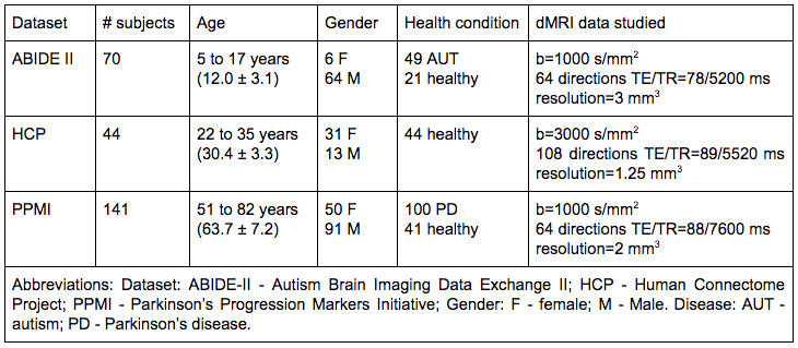
The evaluation dataset (Figure-1; demographics) contained data from a total of 255 subjects across genders, a broad age range (5 to 82 years), health conditions (autism, Parkinson’s disease and healthy subjects), and imaging acquisition protocols (3 different sites). Whole-brain tractography was computed using the two-tensor unscented Kalman filter (UKF) method26,27, which shows high consistency in fiber tracking across different scan protocols and age groups21.
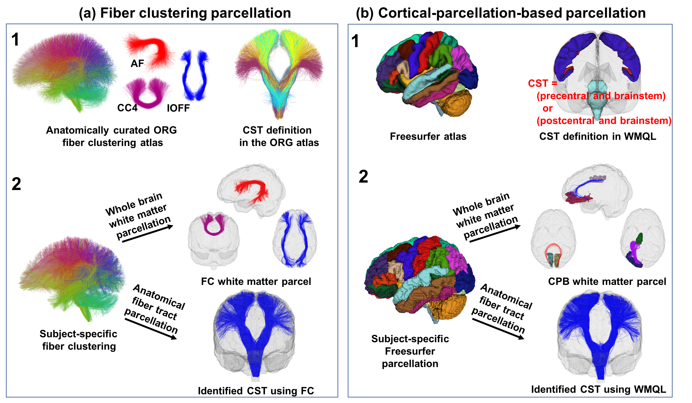
After obtaining tractography, WM parcellation was performed using the FC and CPB methods (Figure-2; method overview). The FC method relied on an O'Donnell Research Group (ORG) fiber clustering atlas21,28 that includes an 800-cluster parcellation of the entire WM and an anatomical fiber tract parcellation. Whole-brain WM parcellation was performed by identifying subject-specific fiber clusters according to the 800-cluster atlas parcellation. Anatomical tract parcellation was performed according to the anatomically curated tracts in the atlas. The CPB method relied on a Freesurfer parcellation atlas to segment an individual’s brain into multiple cortical/subcortical regions. Whole-brain WM parcellation was performed by identifying fiber parcels connecting each pair of the segmented ROIs. Anatomical fiber tract parcellation was performed by leveraging White Matter Query Language (WMQL)29, which provides anatomical definitions of fiber tracts based on their intersected Freesurfer regions.
We computed test-retest measurements of the parcellated WM structures (whole-brain parcels or anatomical tracts) to evaluate the parcellation reproducibility. We computed the weighted Dice (wDice) coefficient3 to measure volumetric overlap of fiber tracts, and relative difference (RD) of fractional anisotropy (FA) to assess the reproducibility of the mean FA of the voxels where the parcellated WM structures were located. A higher wDice and lower RD of FA indicate greater reproducibility.
RESULTS
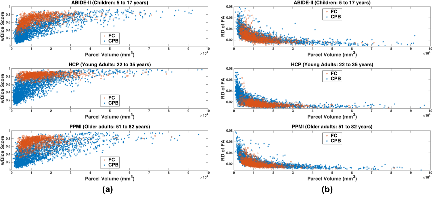
Comparison using all parcels in whole-brain WM parcellation: Significantly higher mean wDice was obtained using FC compared to CPB, with p<0.001 (unpaired t-test, two-tailed) for all of the three datasets (Figure-3a). Significantly lower mean RD was obtained using FC compared to CPB, with p<0.001 (unpaired t-test, two-tailed) for all of the three datasets (Figure-3b).
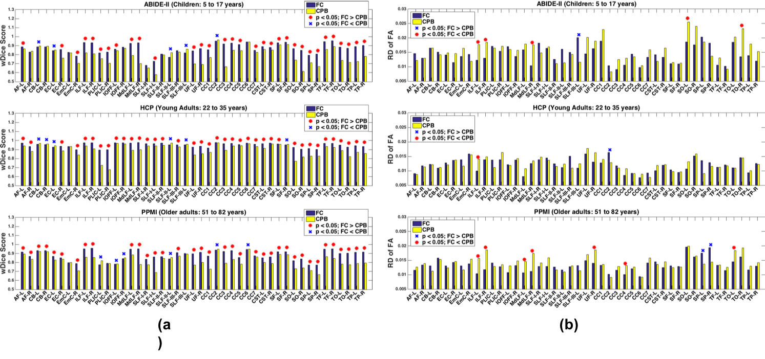
Comparison using 45 individual anatomical tracts: On average across the 3 datasets, using the FC method wDice was significantly higher (p<0.05, paired t-test, two-tailed; FDR corrected) in 73.10% of tracts, while using the CPB method wDice was significantly higher in 12.59% of tracts (Figure-4a). Using the FC method, RD of FA was significantly lower (p<0.05, paired t-test, two-tailed; FDR corrected) in 9.63% of tracts, while using the CPB method RD of FA was significantly lower in 2.96% of tracts (Figure-4b).
DISCUSSION
Overall, the FC method had significantly higher reproducibility than the CPB method in WM parcellations for dividing the entire WM and identifying anatomical fiber tracts. In related work on WM parcellation test-retest reproducibility, good thresholds of wDice and mean RD were considered to be 0.723 and 0.0127. Compared to these findings, we found that both of the FC and CPB methods in the present study performed relatively well, given that 99.26% of CPB tracts and 87.41% of FC tracts had mean wDice scores over 0.72, and the mean RD values of FC tracts and CPB tracts were 0.0122 and 0.0139, which were near 0.012.CONCLUSION
We assessed test-retest reproducibility of two popular WM parcellation strategies, including a white-matter-atlas-based FC method and a Freesurfer-based CPB method. Our experimental results in general indicate that the FC method produced more reproducible WM parcellations than the CPB method.Acknowledgements
We gratefully acknowledge funding provided by the following National Institutes of Health (NIH) grants: P41 EB015902, P41 EB015898, R01 MH074794, R01 MH097979, U01 CA199459, and R03 NS088301.References
1. O’Donnell LJ, Golby AJ, Westin C-F. Fiber clustering versus the parcellation-based connectome. Neuroimage. 2013;80:283-289.
2. Besseling RMH, Jansen JFA, Overvliet GM, et al. Tract specific reproducibility of tractography based morphology and diffusion metrics. PLoS One. 2012;7(4):e34125.
3. Cousineau M, Jodoin P-M, Morency FC, et al. A test-retest study on Parkinson’s PPMI dataset yields statistically significant white matter fascicles. Neuroimage Clin. 2017;16:222-233.
4. Heiervang E, Behrens TEJ, Mackay CE, Robson MD, Johansen-Berg H. Between session reproducibility and between subject variability of diffusion MR and tractography measures. Neuroimage. 2006;33(3):867-877.
5. Kristo G, Leemans A, de Gelder B, Raemaekers M, Rutten G-J, Ramsey N. Reliability of the corticospinal tract and arcuate fasciculus reconstructed with DTI-based tractography: implications for clinical practice. Eur Radiol. 2013;23(1):28-36.
6. Lin C-C, Tsai M-Y, Lo Y-C, et al. Reproducibility of corticospinal diffusion tensor tractography in normal subjects and hemiparetic stroke patients. Eur J Radiol. 2013;82(10):e610-e616.
7. Papinutto ND, Maule F, Jovicich J. Reproducibility and biases in high field brain diffusion MRI: An evaluation of acquisition and analysis variables. Magn Reson Imaging. 2013;31(6):827-839.
8. Tensaouti F, Lahlou I, Clarisse P, Lotterie JA, Berry I. Quantitative and reproducibility study of four tractography algorithms used in clinical routine. J Magn Reson Imaging. 2011;34(1):165-172.
9. Wang JY, Abdi H, Bakhadirov K, Diaz-Arrastia R, Devous MD Sr. A comprehensive reliability assessment of quantitative diffusion tensor tractography. Neuroimage. 2012;60(2):1127-1138.
10. Yendiki A, Reuter M, Wilkens P, Rosas HD, Fischl B. Joint reconstruction of white-matter pathways from longitudinal diffusion MRI data with anatomical priors. Neuroimage. 2016;127:277-286.
11. Besson P, Lopes R, Leclerc X, Derambure P, Tyvaert L. Intra-subject reliability of the high-resolution whole-brain structural connectome. Neuroimage. 2014;102 Pt 2:283-293.
12. Bonilha L, Gleichgerrcht E, Fridriksson J, et al. Reproducibility of the Structural Brain Connectome Derived from Diffusion Tensor Imaging. PLoS One. 2015;10(8):e0135247.
13. Buchanan CR, Pernet CR, Gorgolewski KJ, Storkey AJ, Bastin ME. Test–retest reliability of structural brain networks from diffusion MRI. Neuroimage. 2014;86:231-243.
14. Dennis EL, Jahanshad N, McMahon KL, et al. Development of brain structural connectivity between ages 12 and 30: a 4-Tesla diffusion imaging study in 439 adolescents and adults. Neuroimage. 2013;64:671-684.
15. Duda JT, Cook PA, Gee JC. Reproducibility of graph metrics of human brain structural networks. Front Neuroinform. 2014;8:46.
16. Schumacher LV, Reisert M, Nitschke K, et al. Probing the reproducibility of quantitative estimates of structural connectivity derived from global tractography. Neuroimage. 2018;175:215-229.
17. Smith RE, Tournier J-D, Calamante F, Connelly A. The effects of SIFT on the reproducibility and biological accuracy of the structural connectome. Neuroimage. 2015;104:253-265.
18. Vaessen MJ, Hofman PAM, Tijssen HN, Aldenkamp AP, Jansen JFA, Backes WH. The effect and reproducibility of different clinical DTI gradient sets on small world brain connectivity measures. Neuroimage. 2010;51(3):1106-1116.
19. Zhang Z, Descoteaux M, Zhang J, et al. Mapping population-based structural connectomes. Neuroimage. 2018;172:130-145.
20. Zhao T, Duan F, Liao X, et al. Test-retest reliability of white matter structural brain networks: a multiband diffusion MRI study. Front Hum Neurosci. 2015;9:59.
21. Zhang F, Wu Y, Norton I, et al. An anatomically curated fiber clustering white matter atlas for consistent white matter tract parcellation across the lifespan. Neuroimage. 2018;179:429-447.
22. Fischl B. FreeSurfer. Neuroimage. 2012;62(2):774-781.
23. Di Martino A, O’Connor D, Chen B, et al. Enhancing studies of the connectome in autism using the autism brain imaging data exchange II. Sci Data. 2017;4:170010.
24. Van Essen DC, Smith SM, Barch DM, et al. The WU-Minn Human Connectome Project: an overview. Neuroimage. 2013;80:62-79.
25. Marek K, Jennings D, Lasch S, et al. The Parkinson Progression Marker Initiative (PPMI). Prog Neurobiol. 2011;95(4):629-635.
26. Malcolm JG, Shenton ME, Rathi Y. Filtered multitensor tractography. IEEE Trans Med Imaging. 2010;29(9):1664-1675.
27. Reddy CP, Rathi Y. Joint Multi-Fiber NODDI Parameter Estimation and Tractography Using the Unscented Information Filter. Front Neurosci. 2016;10:166.
28. http://dmri.slicer.org/atlases
29. Wassermann D, Makris N, Rathi Y, et al. The white matter query language: a novel approach for describing human white matter anatomy. Brain Struct Funct. 2016;221(9):4705-4721.
30. Norton I, Essayed WI, Zhang F, et al. SlicerDMRI: Open Source Diffusion MRI Software for Brain Cancer Research. Cancer Res. 2017;77(21):e101-e103.
Figures