3360
Phase-Error-Free Multi-shot 3D DWI using Filtered Back Projection Reconstruction1AllTech Medical Systems, Chengdu, China, 2Alltech Medical System America, Solon, OH, United States
Synopsis
Conventional multi-shot DWI is known to suffer from inter-shot phase inconsistencies due to motion and hardware imperfections. In this work, we present a new approach for phase-error-free diffusion imaging by using multi-shot Paddlewheel-shaped EPI acquisition and filtered back projection (FBP) Reconstruction. The necessity of inter-shot phase correction is removed due to the magnitude-only nature of FBP. Reduced FOV excitation scheme is incorporated to reduce scan time and artifacts. Exemplary results of head and prostate DWI are demonstrated to show the efficacy of the proposed methods.
Introduction
Multi-shot interleaved EPI or spiral trajectories are widely used for high-resolution 3D DWI. But these approaches often suffer from inter-shot phase inconsistencies due to subtle motion or hardware imperfections, which can be difficult to stably correct and hence lead to image blurring and signal loss1-9. Multiplexed sensitivity encoding (MUSE)2-5 or navigator based approaches6-9 can be used to fully calibrate the phase inconsistencies among different shots. However, they are highly challenging for 3D cases because MUSE may be hampered by the noise amplification effects for a large number of shots2-5 and navigator-based approaches cannot easily solve the distortion mismatch between the navigator echo and imaging echo8-9. In this work, a paddlewheel-shaped EPI readout sampling is developed with stack-of-stars radial acquisition. The data from each shot can be first Fourier transformed to form a projection plane. All the planes can then be combined and reconstructed via 2D filtered back projection (FBP) with trivial computational cost. The necessity of inter-shot phase correction is thus removed due to the magnitude-only nature of FBP, making it immune to phase errors.Theory and Method
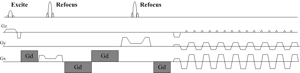
Acquisition: a 3D Planes-on-a-Paddlewheel (POP) EPI readout sampling (Fig 1) was developed for stack-of-stars radial acquisition. During each shot, the phase encoding blips were placed on the slice direction and the sampling trajectory traversed in a standard Cartesian EPI manner. The data acquisition plane then rotated along kz axis until the whole 3D k-space was filled. The double refocus method with bipolar diffusion gradients10 was used to minimize the eddy currents induced by the diffusion gradients. EPI reference scans were also acquired at each azimuthal angle to correct errors caused by gradient delays, B0 inhomogeneity and eddy currents.
Reduced FOV: A reduced FOV excitation scheme was also incorporated to improve radial sampling efficiency. The slice selection gradient lobes were placed at three different axes to excite a volume. Crusher gradients were applied to spoil any unwanted signals.
Gradient Delay: Gradient delays were measured on a sphere phantom via Peters’ method11.The radial trajectories were corrected for gradient delays by addition a constant timing compensation for Gx & Gy.
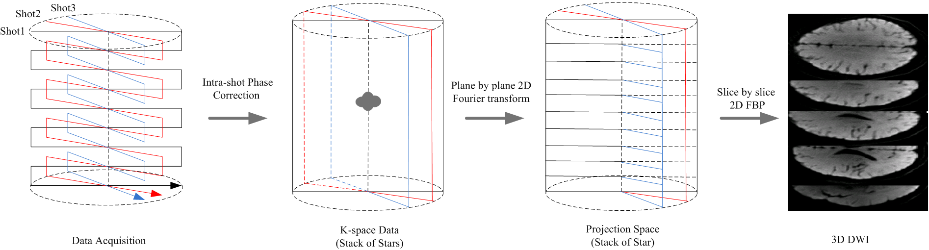
Reconstruction: Fig 2 shows the reconstruction flow. First, the reference scan data were used to perform intra-shot phase correction as a standard EPI pre-processing procedure. The data from each shot was then Fourier transformed into the projection space perpendicular to the acquisition plane. Finally, 2D FBP was used to transform the projection data back to image domain for each slice. The whole reconstruction process was performed on a PC with Inter i7@3.6GHz and 32GB RAM using Matlab.
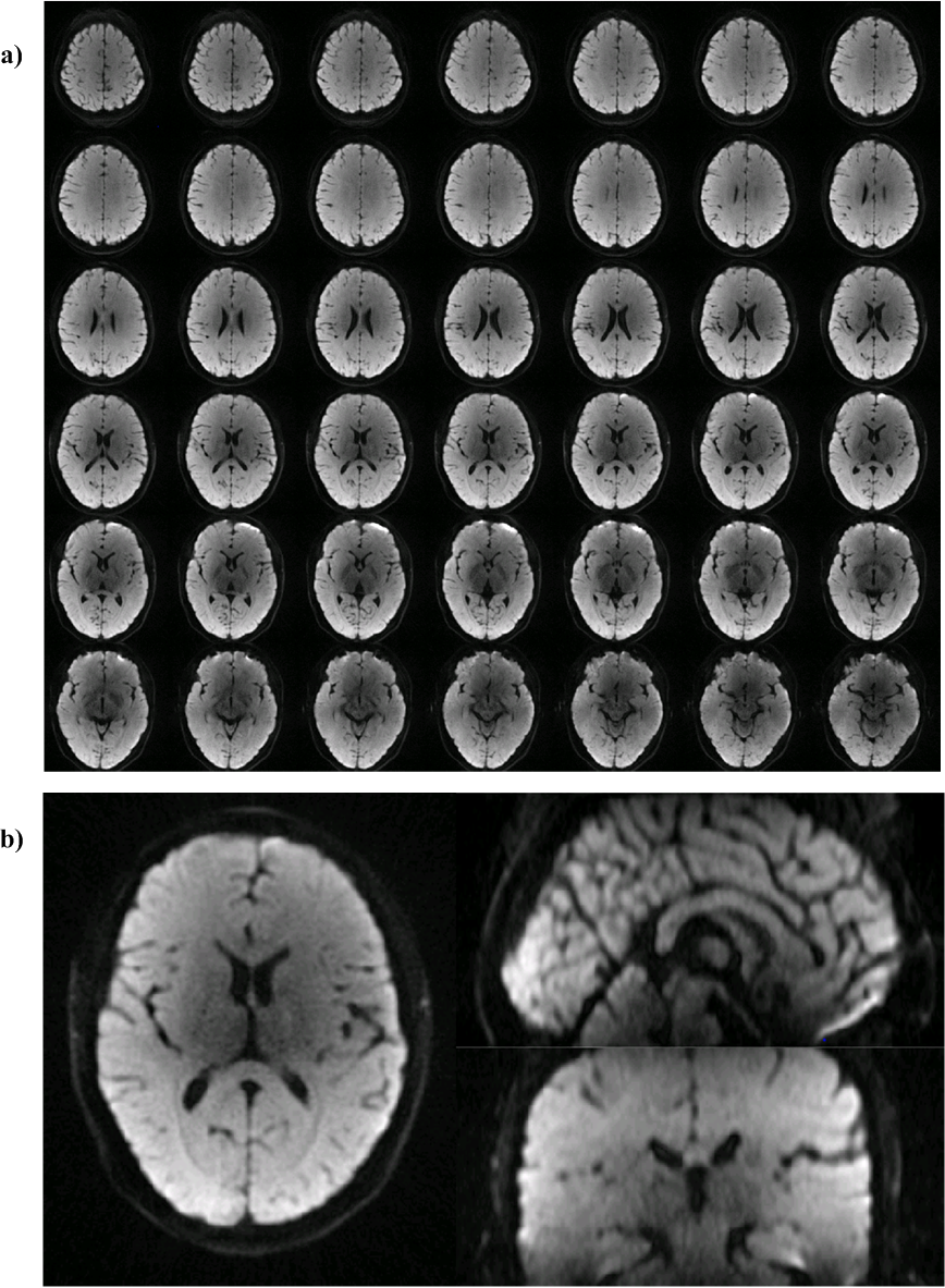
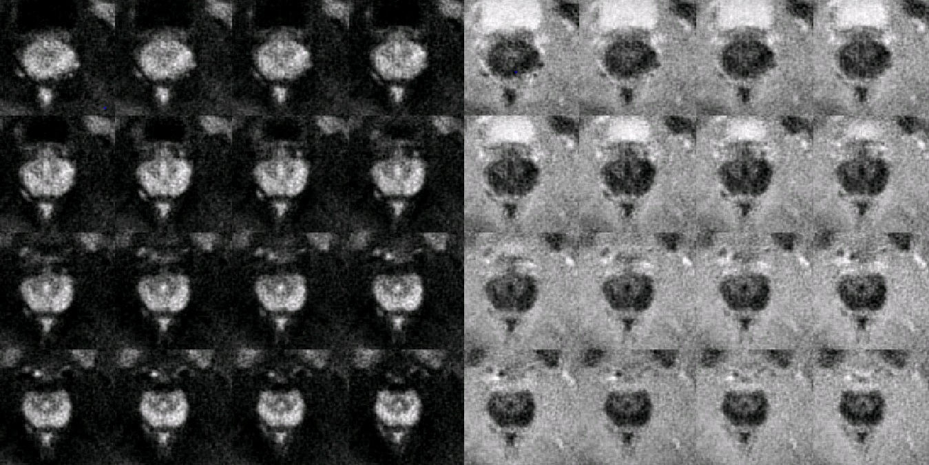
The proposed method was implemented on a 1.5T whole-body scanner (Centauri, Alltech Medical Systems), with gradient strength 33mT/m and slew rate 130mT/m/s. Head and prostate DWI data were acquired. Head DWI parameters: FOV 200*200*90mm3, 10% slice-oversample, 1.3mm isotropic resolution, 3 diffusion directions, b value 0/800, 240 spokes per stack, FA/TR/TE 60/500ms/77.6ms, NEX 1 and total scan time 600s. Prostate DWI parameters: FOV 100*100*60mm3, 10% slice-oversample, 1mm isotropic resolution, 3 diffusion directions, b value 0/800, 150 spokes per stack, FA/TR/TE 60/500ms/75.6ms, NEX 1 for b0, NEX 2 for b800 and total scan time 600s.
Results
Head DWI results from a healthy volunteer is demonstrated in Fig 3. No visible signal corruption, artifact or noise amplification as a result of inter-shot phase variations can be seen. The Image intensity inhomogeneity is due to the uncorrected phase array coil sensitivity variation.
Fig 4. Shows representative results of the prostate DWI and ADC map from a healthy volunteer. Reducing the FOV lead to decreased scan time and enabled high-resolution acquisition of the prostate without visible susceptibility or motion artifacts.
Discussion
The proposed approach can provide robust, high SNR 3D diffusion imaging without any inter-shot phase inconsistency issues, which mainly benefits from the radial sampling scheme and FBP reconstruction. Compared to the conventional multi-shot DWI techniques that require complex inter-shot phase correction mechanisms, the computational cost is trivial. In our case, the reconstruction time for a matrix size of 256*256*80 is less than 10 second via Matlab with single thread.
Slight blurring can be observed in the results, which we suspect is due to the inaccurate radial trajectories. This is an inherent problem in radial imaging, and in particular with the EPI readout scheme. Efforts will be made to further improve the sampling trajectory accuracies in the future work.
Radial sampling is inherently less efficient than Cartesian sampling. With the reduced FOV excitation, the sampling efficiency is improved but the total scan time is still long. Further scan acceleration can be achieved by incorporating model-based reconstruction techniques like compressed sensing.
Acknowledgements
No acknowledgement found.References
- Van AT, Hernando D, Sutton BP. Motion-induced phase error estimation and correction in 3D diffusion tensor imaging. IEEE Trans Med maging 2011, 30:1933–1940.
- Chen NK, Guidon A, Chang HC, Song AW. A robust multi-shot scan strategy for high-resolution diffusion weighted MRI enabled by multiplexed sensitivity-encoding (MUSE). NeuroImage 2013, 72:41–47.
- Chu ML, Chang HC, Chung HW, Truong TK, Bashir MR, Chen NK. POCS-based reconstruction of multiplexed sensitivity encoded MRI (POCSMUSE): a general algorithm for reducing motion-related artifacts. Magn Reson Med 2015, 74:1336–1348.
- Zhang Z, Huang F, Ma X, Xie S, Guo H. Self-feeding MUSE: a robust method for high resolution diffusion imaging using interleaved EPI. NeuroImage 2015;105:552–560.5.
- Chang HC, Hui ES, Chiu PW, Liu X, Chen NK. Phase correction for three-dimensional (3D) diffusion-weighted interleaved EPI using 3D multiplexed sensitivity encoding and reconstruction (3D-MUSER). Magn Reson Med 2018, 79:2702–27126.
- Engstrom M, Skare S. Diffusion-weighted 3D multislab echo planar imaging for high signal-to-noise ratio efficiency and isotropic image resolution. Magn Reson Med 2013, 70:1507-1514.
- Wu W, Poser BA, Douaud G, Frost R, In MH, Speck O, Koopmans PJ, Miller KL. High-resolution diffusion MRI at 7T using a threedimensional multi-slab acquisition. Neuroimage 2016, 143:1–14.
- Dai EP, Ma XD, Zhang Z, Yuan C, and Guo H. Simultaneous Multi-slice Accelerated Interleaved EPI DWI Using Generalized Blipped-CAIPI Acquisition and 3D K-Space Reconstruction. Magn Reson Med 2017, 77:1593–1605.
- Zhang QW, Coolen BF, Nederveen AJ and Strijkers GJ. Three‐dimensional Diffusion Imaging with SPiral Encoded Navigators from Stimulated Echoes (3D‐DISPENSE). Magn Reson Med 2018, DOI: 10.1002/mrm.27470.
- Reese TG, Heid O, Weisskoff RM, and Wedeen VJ. Reduction of Eddy-Current-Induced Distortion in Diffusion MRI Using a Twice-Refocused Spin Echo. Magn Reson Med 2003, 49:177–182.
- Peters DC, Andrew DJ, and McVeigh ER. Centering the Projection Reconstruction Trajectory: Reducing Gradient Delay Errors. Magn Reson Med, 50:1–6 (2003)
Figures