3356
Fast reconstruction of fractional anisotropy with two-dimensional principal component analysis based recognition1The Key Laboratory for Interdisciplinary Research, Institute of Biophysics, China Academy of Sciences, Beijing, China, 2State Key Laboratory of Brain and Cognitive Science, Institute of Biophysics, Chinese Academy of Sciences, Beijing, China, 3The Innovation Center of Excellence on Brain Science, Chinese Academy of Sciences, Beijing, China, 4University of Chinese Academy of Sciences, Beijing, China, 5Siemens Shenzhen Magnetic Resonance Ltd., Shenzhen, China, 6Department of Radiology and Biomedical Imaging, University of California San Francisco, San Francisco, CA, United States, 7UCSF/UC Berkeley Joint Graduate Group in Bioengineering San Francisco, San Francisco, CA, United States
Synopsis
Reducing the acquisition time for obtaining fractional anisotropy (FA) is of paramount importance to investigate cerebral microstructures and morphologies non-invasively. This is the first time to introduce the two-dimensional principal component analysis recognition reconstruction (i.e. 2D-PCA-RR) in recovering highly under-sampled FA maps with 5-fold acceleration of data acquisition. An in-house data processing procedure is implemented to optimize signal-to-noise ratio and construct a distortion-free database. Our results from two different under-sampling patterns show a superior performance gain from the 2D-PCA-RR algorithm as compared to conventional reconstruction methods.
Introduction
Fractional anisotropy (FA) quantitatively characterizes orientation dependence of molecular mobility, which has been widely studied in anatomical biology and medicine [1]. It is usually measured by diffusion tensor imaging (DTI) which is known as a time-consuming approach [2]. One may use parallel imaging for acceleration supported by dedicated hardwares [3]. Alternatively, randomly under-sampling technique has shown its great capability in reducing the scale of data needed for accurate post-processing, either using sparse transformation or pattern recognition [4,5]. Two-dimensional principal component analysis (2D-PCA) has been demonstrated feasible and fast convergent in reconstructing highly under-sampled T1 weighted images, which was named as 2D-PCA-RR [6].
Here, we adopted the 2D-PCA-RR algorithm to reconstruct the FA maps from highly under-sampling DTI datasets obtained at a magnetic field as high as 7 Tesla. An in-house DTI processing pipeline was implemented to attain maximal signal-to-noise ratio (SNR) and correct distortions. The new approach was validated in two cases with different under-sampling patterns. The performance of the approach was compared with conventional reconstruction methods and evaluated by calculating the peak signal-to-noise ratio (PSNR).
Methods
Database construction
Twenty subjects’ DTI datasets were used to construct the database as an input for the 2D-PCA algorithm. The study was approved by the institutional review board. Informed consent was obtained from all participants prior to the scans. All MRI experiments were performed on a 7T research scanner (Siemens Healthcare, Erlangen, Germany) equipped with a 1Tx/32Rx coil. A fully-sampled spin-echo echo planar imaging (EPI) was acquired for the diffusion dataset on each subject (1 mm isotropic resolution, TR = 7 s, TE = 55 ms, matrix = 180x180x102). One frame with b=0 and 30 frames along different diffusion directions with b= 700 s/mm2 were obtained in each dataset. Subsequently, a reverse phase encoding DTI dataset was acquired for distortion correction. The total acquisition time for the DTI study was 21 minutes for each subject.
Fully sampled DTI data processing procedure
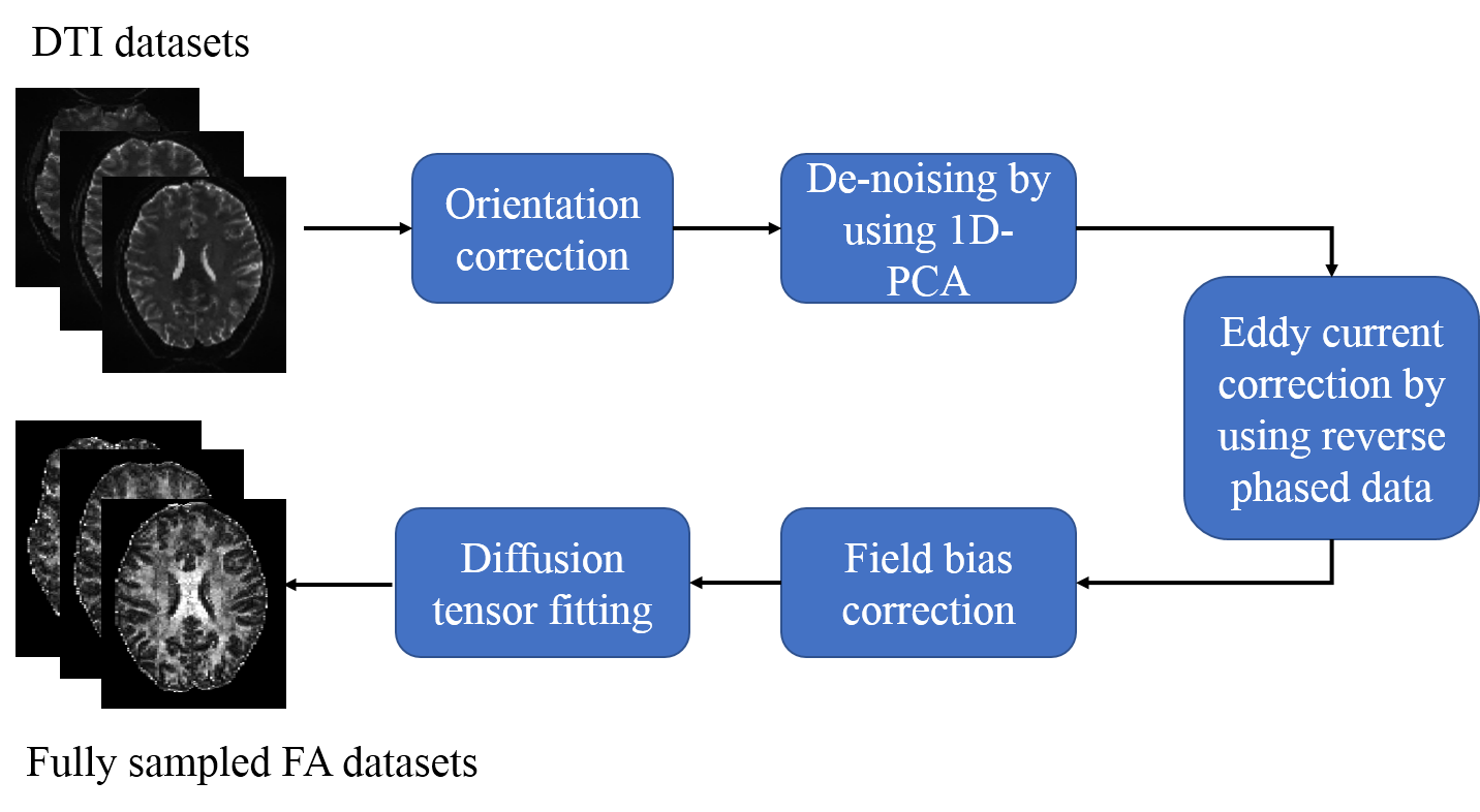
After acquisition, the dataset was visually checked to verify that the images were free from major artifacts. Fully-sampled DTI signal processing followed an in-house pipeline as illustrated in FIG.1, which corrects the distortions, such as eddy-current, subject head movement and field inhomogeneity effects. Fully-sampled FA maps were then be used to build the distortion free database for the 2D-PCA-RR algorithm.
The 2D-PCA-RR algorithm
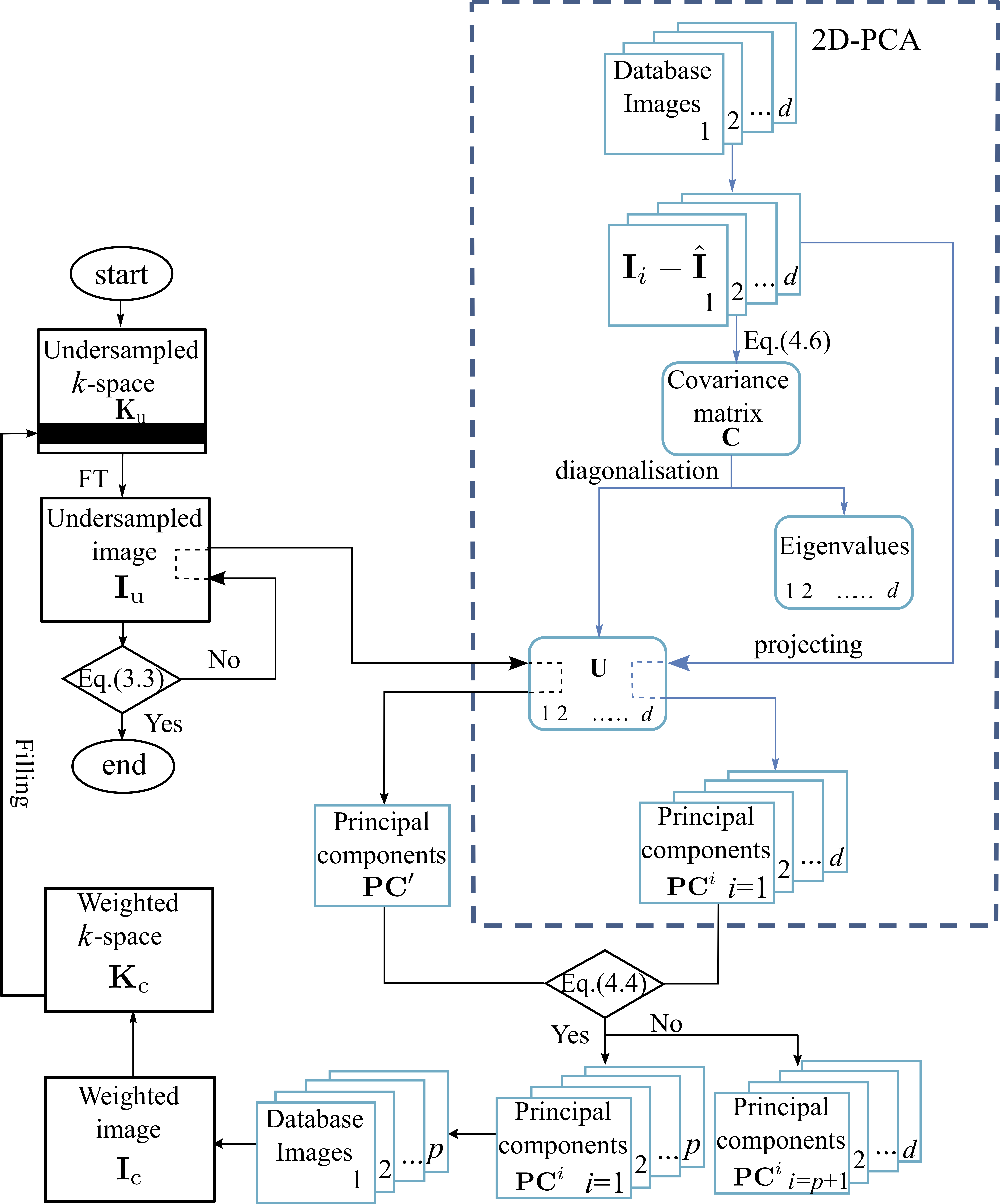
The 2D-PCA-RR algorithm approximates the under-sampled image by iteratively improving estimations for the missing k-space data. We adopted the algorithm for the FA reconstruction which is detailed in FIG.2. This approach is based on the recognition of an averaged image originating from the fully-sampled FA database to the initially under-sampled information as input. This procedure is performed in a loop until the improvement in the reconstructed image falls below a preset threshold for subsequent iterations. All algorithms were implemented in MATLAB 2015a (The Mathworks, Natick, MA).
Results and discussions
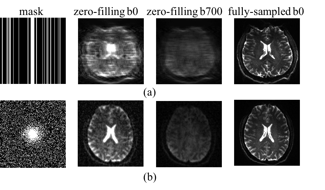
FIG.3 shows the two under-sampled cases generated from the fully-sampled DTI datasets at different b values. The first case was to randomly sample 20 percent phase encoding lines. In order to explore the feasibility of extending the proposed method to other k-space trajectories, the second case was simulated by applying the undersampling on a spiral acquisition with the same sampling rate. As observed from FIG.3, the under-sampling in the EPI trajectory induces more artifacts than the spiral under-sampling, indicating that the under-sampling in EPI sequence poses more challenges to the reconstruction algorithm.
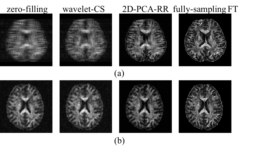
The reconstructed FA maps by using the 2D-PCA-RR algorithm were compared with the zero-filling and sparse transformation (wavelet compressed sensing, i.e. wavelet-CS) reconstruction and shown in FIG.4. The PSNR of the three reconstruction methods were 16.26 (zero-filling), 19.63 (wavelet-CS), 21.93 (2D-PCA-RR) in the case of EPI-under-sampling, and 15.80 (zero-filling), 20.76 (wavelet-CS), 23.74 (2D-PCA-RR) in the case of spiral under-sampling, respectively. The 2D-PCA-RR results show more details in both cases, which demonstrate its feasibility in the reconstruction of FA maps. As compared to the spiral under-sampling, the proposed approach has a superior performance gain in randomly under-sampling the EPI trajectory, which is widely used in DTI acquisition.
Conclusions
This is the first study to reconstruct highly under-sampled FA maps by using 2D-PCA-RR with 5-fold acceleration of the DTI acquisition. Our findings demonstrate that 2D-PCA-RR has unique capabilities in accurately recovering FA maps, either originating from EPI or spiral under-sampling. Moreover, this approach may be extended to reconstruct other diffusion matrices, such as diffusion kurtosis which takes even longer acquisition time than FA.Acknowledgements
We thank the Ministry of Science and Technology of China via the grant 2015CB351701, the National Nature Science Foundation of China via the grant 91132302 and the Chinese Academy of Sciences via the Hundred Talents Program Class-A Award for financially supporting this study.
References
[1] Basser P J, Inferring microstructural features and the physiological state of tissues from diffusion-weighted images. NMR Biomed 1995;8:333–344
[2] Basser P J, Mattiello J, LeBihan D, MR diffusion tensor spectroscopy and imaging. Biophys J 1994;66:259–267
[3] Hutchinson M, Raff U. Fast MRI data acquisition using multiple detectors. Magn Reson Med 1988;6:87–91.
[4] Lustig M, Donoho DL, Sparse Pauly JM, MRI. The application of compressed sensing for rapid MR imaging. Magn Reson Med 2007;58:1182–1195.
[5] Zong FR, d'Eurydice MN, Galvosas P. Reconstructing undersampled MR Images by utilising principal-component-analysis-based pattern recognition. Proc 12thIBC MRPM Conf, 22(14); 2014. p. 1–5.
[6] Zong FR, d'Eurydice MN, Galvosas P. Fast reconstruction of highly undersampled MR images using one and two dimensional principal component analysis. Magn Reson Imag 2016;34;227–238.
Figures