3355
Pseudo-3D Diffusion-Weighted Imaging of the Brain using Echo Planar Imaging with Compressed SENSE (EPICS)Kosuke Morita1, Masami Yoneyama2, Takeshi Nakaura3, Seitaro Oda3, Masahiro Hatemura1, and Yasuyuki Yamashita3
1Radiology, Kumamoto University, Kumamoto-shi, Japan, 2Philips Japan, Tokyo, Japan, 3Diagnostic Radiology, Kumamoto University, Kumamoto-shi, Japan
Synopsis
We attempted to obtain brain high-resolution pseudo-3D (2D multi-slice acquisition with very thin slice thickness) diffusion-weighted echo planar imaging (DW-EPI) using a hybrid compressed sensing and sensitivity encoding (Compressed SENSE) framework (EPICS). pseudo-3D-DWI with EPICS achieved high-resolution (1.15 mm3) isotropic DWI within clinically feasible scan time. Furthermore, EPICS clearly improved the accuracy and robustness of ADC values in high b-value brain DWI with pseudo-3D acquisition without any penalty for scan parameters.
PURPOSE
Typical clinical brain diffusion-weighted imaging based on Echo Planar Imaging (DW-EPI) images have limited spatial resolution compared to other imaging due to its high sensitivity to B0 inhomogeneities. DWI with smaller voxel size causes further image distortion. DW-EPI with sensitivity encoding (SENSE) helps to reduce the voxel size without increasing of image distortion, but it often suffers from increased noise-like artifacts on the center of the images due to the high geometry factor [1, 2]. Recently, compressed sensing (CS) or Compressed SENSE (C-SENSE) has emerged and it demonstrated that the images from highly undersampled measurements can be reconstructed accurately by using the sparsity of the MR images. We hypothesize that the CS reconstruction can similarly improve image quality drastically for EPI based DWI without further optimization of EPI sampling scheme. It has been shown before that wavelet based denoising is an effective tool for image quality improvement in high b-value DWI images [3, 4], in this work it is integrated with SENSE parallel imaging in an iterative implementation. In this our study, we attempt to obtain brain high-resolution pseudo-3D (p3D, it means 2D multi-slice acquisition with very thin slice thickness) DW-EPI using CS framework (EPICS) and to demonstrate the feasibility of EPICS p3D-DWI.MATERIALS AND METHODS
Experimental data was collected from 5 healthy volunteers. Written informed consent was obtained from each volunteer and the protocol was approved by the ethics committee. All studies were performed with clinical 3.0T MR scanner (Philips, Ingenia 3.0T CX) and 32-channel dS-head coil. EPICS is based single-shot DWI-EPI. We did not modify its sampling pattern of single-shot EPI and we applied it into the C-SENSE framework. Scan parameters of DWI-EPI were as follows: TR / TE = 15174 / 90 ms, field-of-view = 25.6 × 25.6 cm2, acquisition matrix = 224 × 224 (reconstruction matrix = 512 × 512), slices thickness = 1.15 mm, voxel size = 1.14 × 1.14 × 1.15 mm, number of slices = 80, NSA = 6, EPI factor = 55, Band width = 2230.4 Hz, CS factor or SENSE factor = 4.0, b-values = 0 and 1000 sec/mm2, Acquisition time = 6:19-6:25, Transverse plane acquisition. EPICS p3D DWI were compared to conventional SENSE images for image quality, especially for the reduction of image noise. ROIs were placed on left and right white matter (WM). The average signal intensities (SI) of WM on b0 and b1000 image and ADCs of WM were used for comparison between EPICS and SENSE. SIs and ADCs of WM were assessed by using paired t-test.RESULTS
Figure 1 shows results of SIs and ADC value between EPICS and SENSE. There was no significant difference in SI (b0 image). SI (b1000 image) of EPICS and SENSE were 138.03±14.50 and 168.04±30.32 (p < 0.05). ADC values of EPICS (0.72±0.04) was significantly higher than that of SENSE (0.48±0.13) (p < 0.05). The cause of lower ADC values of SENSE was most likely due to the presence of severe noise over the brain. On the other hand, there were no significant differences between ADC values of EPICS and those of reference. Note that the reference ADC values are obtained by conventional clinical DW-EPI sequence we are routinely using in our institute. It indicated that EPICS can provide more accurate ADC values. Figure 2 shows representative b1000 images and ADC maps using EPICS and SENSE in a volunteer. EPICS clearly reduced the noise which exists in the center of the SENSE images. It indicated that EPICS can provide more accurate ADC values with high reproducibility and robustness. Figure 3 shows representative 3D MPR images obtained by p3D-DWI. The p3D-DWI with EPICS achieved high-resolution (1.15 mm3) isotropic DWI within clinically feasible scan time.CONCLUSION
EPICS clearly reduces noise-like artifacts and significantly improves the accuracy and robustness of ADC values in high b-value brain DWI with pseudo-3D acquisition compared with conventional SENSE DW-EPI, without any penalty for scan parameters. This technique may be helpful to further assess the many brain diseases.Acknowledgements
No acknowledgement found.References
[1] Patricia N, et al. Parallel Imaging Artifacts in Body Magnetic Resonance Imaging. Can Assoc Radiol J. 2009;60: 91–98. [2] Yanasak NE, et al. MR imaging artifacts and parallel imaging techniques with calibration scanning: a new twist on old problems. Radiographics. 2014;34:532-48. [3] Wirestam R, et al. Denoising of Complex MRI Data by Wavelet-Domain Filtering: Application to High-b-Value Diffusion-Weighted Imaging. Magn Reson Med. 2006;56:1114-20. [4] Yang X, et al. A wavelet multiscale denoising algorithm for magnetic resonance (MR) images. Meas Sci Technol. 2011;22:025803.Figures
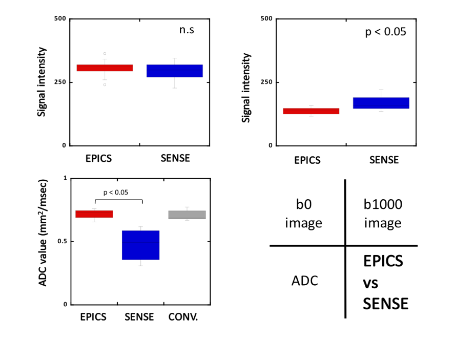
Figure
1. Comparison of signal intensity (b = 0, 1000) and ADC between EPICS and
SENSE.
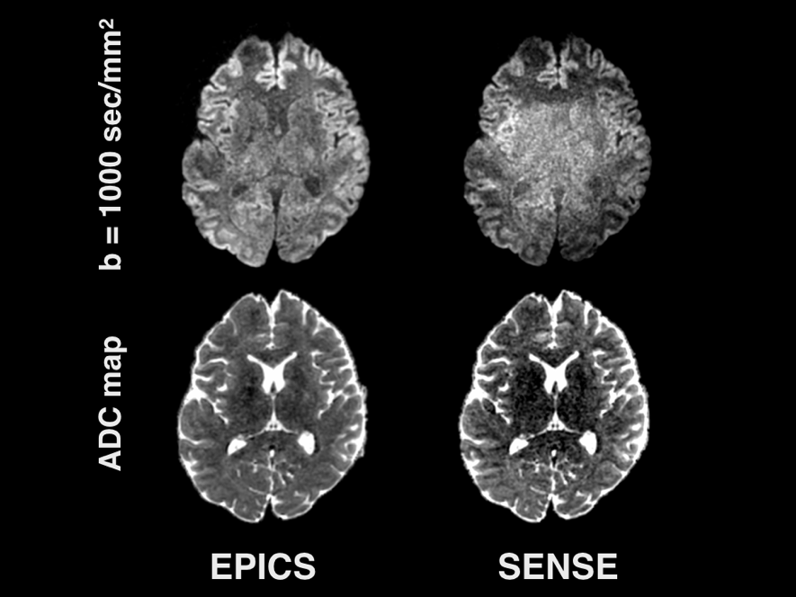
Figure
2. Representative b = 1000 sec/mm2 images and ADC maps using EPICS and SENSE
obese volunteer.
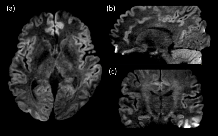
Figure
3. Representative 3D MPR images obtained by EPICS
p3D-DWI (b =
1000 sec/mm2) with high-resolution
isotropic (1.15 mm3) voxel size. Axial source image (a),
sagittal (b) and coronal (c) reformats are shown.