3354
Nyquist Ghost Correction of High-Resolution SMS Breast DWI with Ghost/Object Minimization1Biomedical Engineering, University of Minnesota, Minneapolis, MN, United States, 2Center for Magnetic Resonance Research, University of Minnesota, Minneapolis, MN, United States, 3Radiology, University of Minnesota, Minneapolis, MN, United States
Synopsis
A recent novel approach to acquire high-resolution breast DWI uses a simultaneous multi-slice (SMS) SE-EPI sagittal acquisition. In EPI, Nyquist ghosts are typically corrected using a 3-line navigator, which often fails in SMS SE-EPI breast DWI due to low SNR, insufficient fat suppression, and larger B0 inhomogeneity. In this work we compare a referenceless ghost correction method, called Ghost/Object minimization, with the standard 3-line navigator in high-resolution breast DWI. Ghost/Object provides more reliable 1st-order ghost correction in a dynamic and slice-specific way, which improves image quality and reduces bias in ADC values compared to the standard correction.
Introduction
Diffusion weighted imaging (DWI) is increasingly used in breast cancer imaging because low apparent diffusion coefficients (ADCs) indicate malignancy. However, the ability to detect lesions is extremely limited by the low resolution of typical single-shot spin-echo (SS SE) EPI. We recently developed a simultaneous multi-slice (SMS)[1] approach to SS SE-EPI for breast DWI that was derived from the Human Connectome Project’s (HCP)[2] high-resolution protocol.
In EPI, eddy currents and timing errors cause inconsistencies between the positive and negative readout lines (RO+/RO-) that manifest as Nyquist ghosts in the phase encoding (PE) direction of the image. These ghosts can obstruct the visualization of the DW images and bias ADC values. Ghosts are typically corrected using a 3-line navigator, which typically works reliably in HCP brain imaging, but often fails in high-resolution SMS breast DWI due to insufficient SNR, fat suppression, and B0 homogeneity. Moreover, ghost correction failure in autocalibration scans (ACS) propagates in the SMS and GRAPPA reconstructions that are often employed in high resolution imaging.
Many alternative ghost correction strategies exist, including a referenceless class[3-5], which directly calculate the correction from the data itself. Ghost/Object minimization (G/O)[6] is one referenceless method based in the image domain that was recently proposed in [6]. The purpose of this work is to compare G/O with the standard 3-line navigator ghost correction in high-resolution breast DWI acquired with SMS SE-EPI.
Methods
Acquisition
Sixteen breast cancer patients were scanned prone on a Siemens 3T prismafit with a 16-channel breast coil (Sentinelle) under an IRB-approved protocol. DWI was acquired using 2D SE-EPI with 256 sagittal slices of 1.25 mm with SMS MB=4, which are reformatted to axial images for clinical viewing. The following parameters were used: TR/TE=6500/60.80 ms, 1.25x1.25 mm nominal in-plane resolution, head/foot PE direction, GRAPPA R=2, and monopolar diffusion (4 at b=0, 4 at b=800 s/mm2). Standard T1-weighted anatomical images were also acquired.
Ghost Correction
Two first-order ghost correction methods were applied offline to both the ACS data and each undersampled acquisition on a per-channel and per-slice basis. The 3-line navigator, which represents typical online correction, acquires a navigator through ky=0 (RO+/RO-/RO+) to measure the phase difference. G/O minimization was performed directly on ACS and undersampled data, starting with a wide discrete search and refined with a simplex search. After SMS unaliasing, the ghost correction was adjusted in a slice-specific way, statically for the 3-line navigator (based on the difference between ACS and SMS navigators) and dynamically for G/O (based on a second iteration of the minimization problem).
Analysis
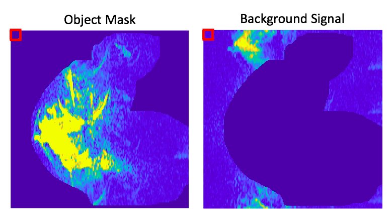
Because ghosts in the background region cause signal change in the object and often imply that some ghost overlaps the object, the mean background signal can act as a surrogate for overall ghost levels. T1-weighted images were automatically masked and resampled onto the DWI to define the object region (Figure 1). The ghost intensity was defined over each volume as the mean background signal compared to the average signal in a noise-only volume (Figure 1).
Results
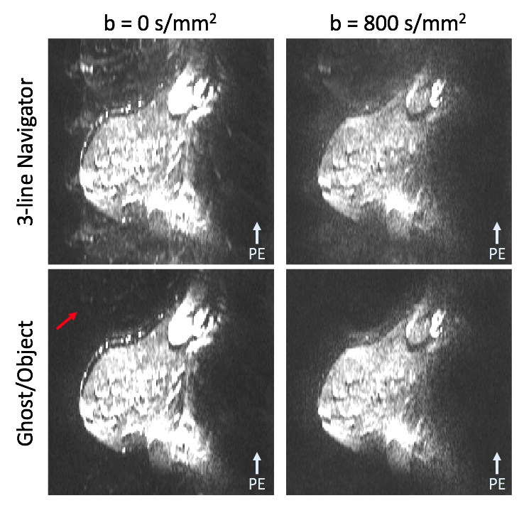
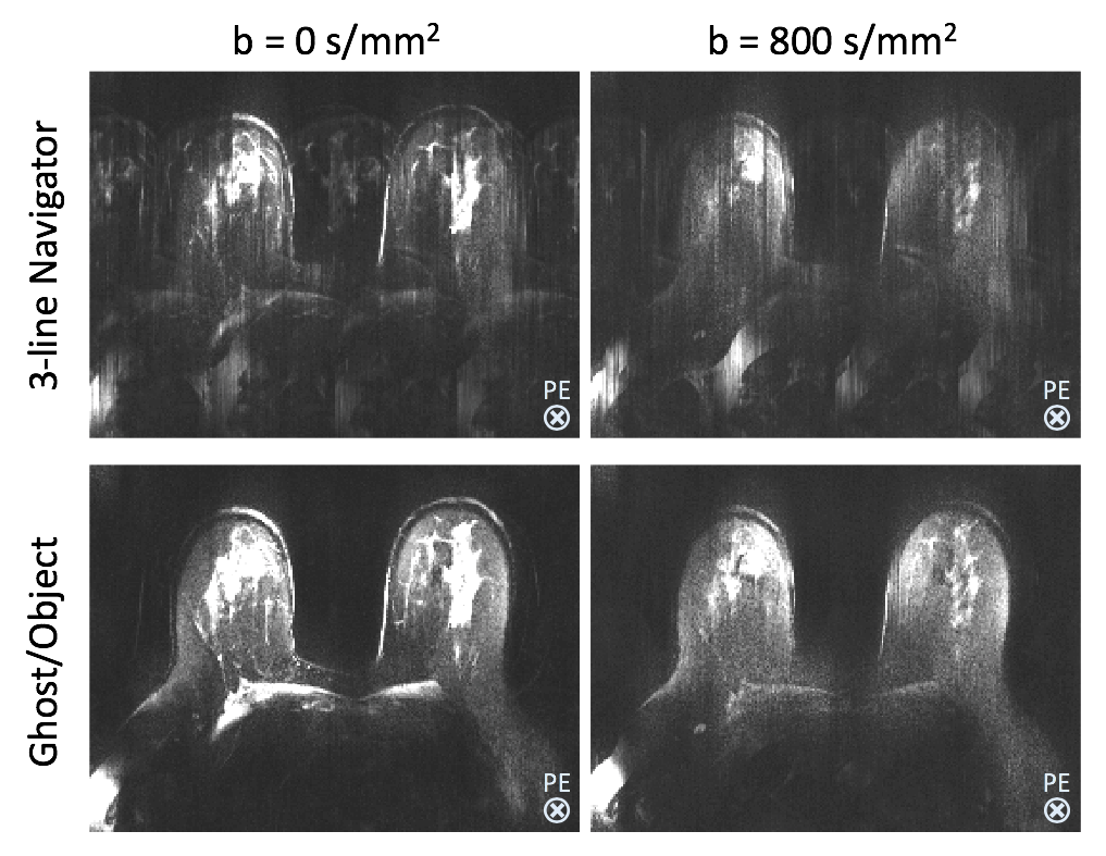
Two examples of in vivo b=0 and 800 s/mm2 images are shown in Figures 2 and 3. In Figure 2, the 3-line navigator clearly fails, causing Nyquist ghosts and poor GRAPPA unaliasing. Small levels of GRAPPA errors are also present in G/O images. Figure 3 demonstrates how the artifacts in the PE direction influence the SMS unaliasing and overall axial image quality. Several artifacts are present in the slice dimension after 3-line navigator correction, which G/O greatly reduces.
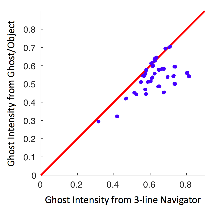
The ghost intensity of G/O is plotted versus the 3-line navigator in Figure 4 for each acquisition. G/O either performs equivalently (on the line) or outperforms (to the right of the line) the standard 3-line navigator for all cases.
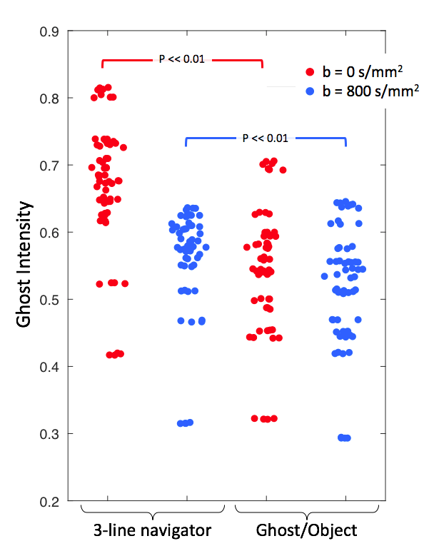
Figure 5 plots ghost intensities separated by b-value. G/O yields significantly lower residual ghost levels than the 3-line navigator at both b-values (paired t-test, p<<0.01, N=16).
Discussion
Nyquist ghosts can be present in either the undersampled data or the ACS reference data, which often causes a failure in GRAPPA reconstruction. Defining the ghost intensity based on the background signal accounts for both Nyquist ghost correction failure and GRAPPA aliasing artifacts.
G/O does not require the acquisition of additional reference data and it can be measured directly from the undersampled data itself, the SMS-unaliased data, and the ACS lines, allowing for a fully slice-specific and dynamic correction.
Conclusions
The standard 3-line navigator is insufficient for ghost correction of high resolution, breast SE-EPI DWI with SMS. The alternative G/O referenceless method provides more reliable 1st-order ghost correction in a dynamic and slice-specific way, which improves image quality and reduces bias in ADC values compared to the standard correction.Acknowledgements
NIH P41 EB015894
NIH R21 CA201834
NIH 1S10 OD017974-01
References
1) Larkman et al. JMRI 2001;13(2)313-7
2) Sotiropoulos et al. NeuroImage. 2013;80:125-45.
3) Clare S. In Proc. 11th Annual ISMRM, Toronto, Ontario, 2003;1041.
4) Skare S, et al. In Proc. 14th Annual ISMRM, Seattle, WA, 2006;2349.
5) Peterson E, et al. In Proc. 23rd Annual ISMRM, Toronto, Ontario, 2015;75.
6) McKay JA, et al. MRM. 2018.
Figures