3352
Whole-brain DTI at 860 μm isotropic resolution in 10 minutes on a commercial 3T Scanner1Ming Hsieh Department of Electrical Engineering, University of Southern California, Los Angeles, CA, United States, 2Martinos Center for Biomedical Imaging, Charlestown, MA, United States
Synopsis
We describe an acquisition and reconstruction methodology that enables in vivo human diffusion tensor imaging with whole-brain coverage and 860$$$\mu$$$m isotropic spatial resolution, all within a 10 minute acquisition window on a commercial 3T scanner. Our approach is enabled by combining the gSlider-SMS acquisition approach (which uses simultaneous multi-slab acquisition for increased spatial coverage, combined with highly-efficient RF slab-encoding to achieve high spatial resolution) with an SNR-enhancing joint reconstruction approach that mitigates the noise associated with high-resolution acquisition.
Introduction
Diffusion tensor imaging (DTI) is a popular method for quantifying brain microstructure and analyzing white matter connectivity, but generally suffers from low-SNR and long data acquisition time. These factors have placed practical limits on the achievable spatial resolution for modern scan protocols.
In this work, we describe an acquisition and reconstruction methodology that enables the collection of 30 diffusion weighted images (DWIs) and 3 unweighted (b=0s/mm$$$^2$$$) images with 860$$$\mu$$$m isotropic spatial resolution and whole-brain coverage, all within a 10 minute acquisition window on a commercial 3T scanner. Our approach is enabled by combining the gSlider-SMS approach for high resolution diffusion imaging1 (which enables high-resolution imaging through simultaneous multi-slab (SMS) acquisition combined with a highly-efficient RF slab-encoding strategy) together with an SNR-enhancing joint reconstruction method2,3 (to mitigate the noise problems resulting from fast high-resolution imaging). SNR-enhancing joint reconstruction has strong theoretical characterizations and makes weaker assumptions than many other alternative SNR-enhancement methods for diffusion MRI data, which can allow it to be more useful than other approaches for images with subtle or highly-localized diffusion features3.
This combination of methods has been previously demonstrated to enable fast high-resolution diffusion imaging on specialized imaging hardware with uncommonly powerful gradients, i.e., the Siemens 3T CONNECTOM system.4,5 The use of this specialized hardware offered a number of benefits, and for example, enabled shorter TE values (and therefore higher SNR) than would be achievable on a more conventional system. In this work, we demonstrate that this same approach can also be used to enable fast diffusion imaging on a commercial 3T scanner with more typical hardware specifications.
Methods
gSlider-SMS was implemented on a Siemens 3T Prisma scanner, and data was acquired from a healthy volunteer with a 32-channel coil. Data was acquired with 860$$$\mu$$$m isotropic resolution across the whole brain, with TE=74ms, TR=3.5s, 3$$$\times$$$ accelerated parallel imaging with an SMS factor of 2 and $$$6/8$$$ths partial Fourier acquistion, 5 different RF slab-encodings per DWI, and 33 total images (30 images with b=1000s/mm$$$^2$$$ + 3 images with b=0s/mm$$$^2$$$). The total acquisition time was slightly under 10 minutes. A B1 map was also acquired to enable compensation for spatially-varying flip angles during RF encoding.
Similar to previous work4,5, parallel imaging and SMS reconstruction was performed in a first step to avoid the need to manipulate extremely large datasets. Subsequently, gSlider reconstruction (which synthesizes a high-resolution image from multiple lower-resolution RF-encoded images1) was performed simultaneously with SNR-enhancing joint reconstruction (leveraging the shared spatial structure possessed by DWIs of the same anatomy, while also enabling strong theoretical characterization and automated tuning of regularization parameters based on desired SNR/resolution trade-offs2-5).
Subsequently, quantitative DTI model parameters were estimated from the reconstruction results. For comparison, we also estimated quantitative DTI model parameters to a gSlider reconstruction of the data without SNR-enhacing joint reconstruction.
Results
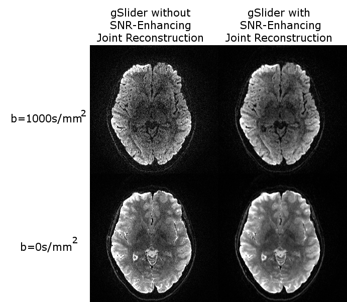
The effects of SNR-enhancing joint reconstruction on the DWIs is illustrated in Fig. 1. As can be seen, the conventional gSlider reconstruction result has very obvious noise contamination, as should be expected given the fast acquisition and high spatial resolution. On the other hand, the SNR-enhancing joint reconstruction result has substantially less noise, and this is achieved without substantially sacrificing the spatial resolution of the images.
The effects of SNR-enhancing joint reconstruction on quantitative DTI parameters is illustrated in Fig. 2. As can be seen, the tensor orientations and FA values estimated from gSlider reconstruction without SNR-enhancing joint reconstruction demonstrate obvious noise artifacts, as should be expected due to the low-SNR observed in Fig. 1. On the other hand, SNR-enhancing joint reconstruction enables high-quality quantitative DTI results.
To further illustrate the information content of this high-resolution data, Fig. 3 shows an overlay of DTI glyphs from a region of the image containing the boundary between white matter and cortical gray matter (visualization performed using BrainSuite, http://brainsuite.org). As can be seen, the spatial resolution is high enough to provide visualization of fine-scale structures, e.g., we have multiple voxels spanning the cortical ribbon. Consistent with the previous figures, this result also demonstrates substantial improvements due to SNR-enhancing joint reconstruction.
Conclusion
Fast high-resolution DTI has previously been difficult to achieve, but is feasible on commercial 3T MRI scanners when using advanced data acquisition and image reconstruction strategies. The substantial improvements to imaging speed and resolution we've achieved have potentially major implications for a wide variety of clinical and neuroscience applications.
Acknowledgements
This work was supported in part by NIH grants R01-MH116173, R01-NS074980, R01-NS089212, and R21-EB022951, NSF grant CCF-1350563, and a USC Viterbi/Graduate School Ph.D.Fellowship.References
1. Setsompop K, Fan Q, Stockmann J, Bilgic B, Huang S, Cauley SF, Nummenmaa A, Wang F, Rathi Y, Witzel T, Wald LL. High‐resolution in vivo diffusion imaging of the human brain with generalized slice dithered enhanced resolution: Simultaneous multislice (gSlider‐SMS). Magn Reson Med 2018;79:141-151.
2. Haldar JP, Wedeen VJ, Nezamzadeh M, Dai G, Weiner MW, Schuff N, Liang Z-P. Improved diffusion imaging through SNR-enhancing joint reconstruction. Magn Reson Med 69:277-289, 2013.
3. Kim JH, Song S-K, Haldar JP. Signal-to-noise ratio-enhancing joint reconstruction for improved diffusion imaging of mouse spinal cord white matter injury. Magn Reson Med 75:1499-1514, 2016.
4. Haldar JP, Fan Q, Setsompop K. Whole-brain quantitative diffusion MRI at 660 µm resolution in 25 minutes using gSlider-SMS and SNR-enhancing joint reconstruction. Proc. ISMRM 2016, p. 102.
5. Haldar JP, Setsompop K. Fast high-resolution diffusion MRI using gSlider-SMS, interlaced subsampling, and SNR-enhancing joint reconstruction. Proc. ISMRM 2017, p. 175.
Figures