3345
Self-navigated Half-Fourier Multi-shot Echo-planar DWI Reconstructions for Brain Imaging1Institute for Signal Processing, University of Lübeck, Lübeck, Germany, 2Philips Research Hamburg, Hamburg, Germany, 3Dept. Radiology, LUMC, Leiden, Netherlands
Synopsis
EPI trajectories using Half-Fourier achieve shorter echo times and therefore higher SNR, which is especially desirable in low-SNR applications like diffusion-weighted MRI. For the same reason, methods enabling phase-corrected image recovery for multi-shot diffusion acquisitions have been intensively studied for both spiral and EPI trajectories. In this work, two algorithms are presented comprising both half-Fourier and the multi-shot same-magnitude constraints to exploit the advantages of both techniques. The algorithms are shown to robustly recover interleaved half-Fourier datasets from in-vivo brain acquisitions.
Introduction
Single-shot EPI is currently the standard acquisition scheme in diffusion-weighted MRI (DWI)1. Recently, several SENSE-based2,3 multi-shot reconstruction algorithms like POCS-ICE4 and MUSE5 have been proposed for spiral and Echo-planar Imaging (EPI) trajectories, respectively, to increase SNR. Besides, half-Fourier (HF) is another common acceleration strategy6 to reduce echo times and further improve SNR by reducing $$$T_2$$$ -decay and –shine-through. This work presents two novel DWI algorithms for interleaved half-Fourier EPI reconstructions. To the best of our knowledge, this combined problem has not yet been addressed in the literature.Theory
POCS-ICE is an iterative SENSE-based algorithm for multi-shot spiral DWI comprising five basic steps4. First, data projection is performed in k-space. Then, shots are reconstructed using SENSE. Next, motion-induced shot phases are estimated followed by phase-corrected shot combination. Finally, the next shot guesses are formed by recombining the joint image and the shot-specific phase.
Half-Fourier techniques assume the complex image to be real-valued except for a low-resolution phase6. Appropriate reconstruction schemes therefore comprise two main steps. Firstly, the low-resolution phase is estimated from the fully-sampled k-space center. Secondly, missing k-space data is recovered by conjugate symmetry. In practice, POCS-based schemes are used iteratively enforcing data consistency in k-space and phase consistency in image space
Methods
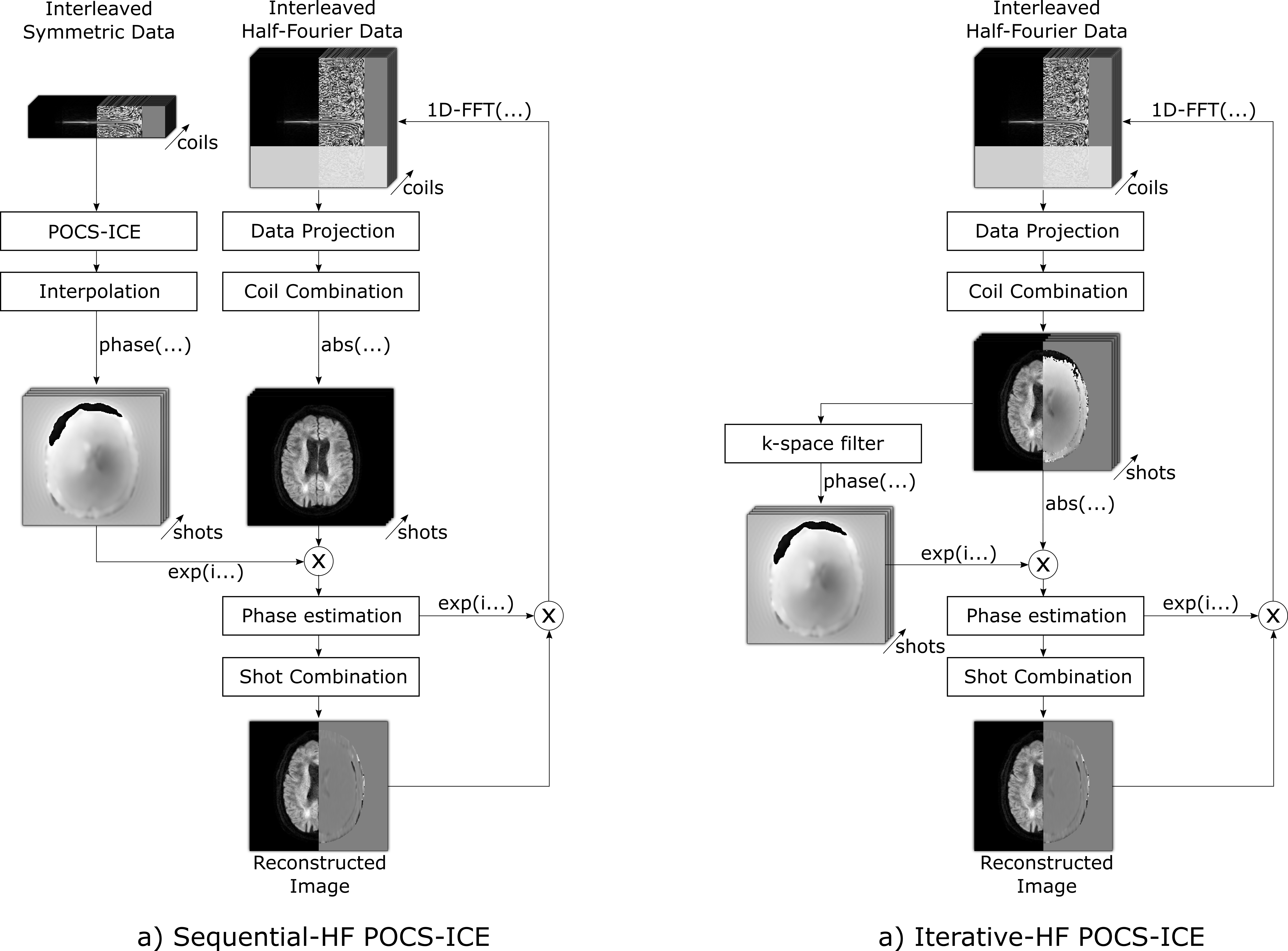
The algorithms in this work extend POCS-ICE by the half-Fourier phase consistency constraint. After data projection and coil combination, half-Fourier phase projection7 is performed replacing the shot phases by half-Fourier low-resolution phase estimates. Next, conventional shot combination is performed using a 2D triangular window4 with the window size set to the available range of the symmetric data. Two algorithms were tested for the aforementioned half-Fourier phase estimation. The structures are schematically illustrated in Figure 1.
1) Sequential-HF POCS-ICE does the half-Fourier phase estimation once in advance. Firstly, the symmetric data is reconstructed by conventional POCS-ICE4, interpolated by zero-filling in k-space, additional 2D-Hann-window-filtering adapted to the symmetric matrix size and phase extraction (no unwrapping).
2) Iterative-HF POCS-ICE does the half-Fourier phase estimation within each iteration. The shot images are therefore filtered in k-space by 2D-Hann-window-filtering adapted to the symmetric matrix size and subsequent phase extraction (no unwrapping).
The Cartesian EPI undersampling properties were exploited to accelerate computations. For the HF reconstruction, data projections are performed in hybrid $$$x$$$-$$$k_y$$$-space replacing 2D- by 1D-FFTs7. For the symmetric part, the undersampling can be described in image-space using the point spread function2 and phase ramps for the shot trajectory shifts.
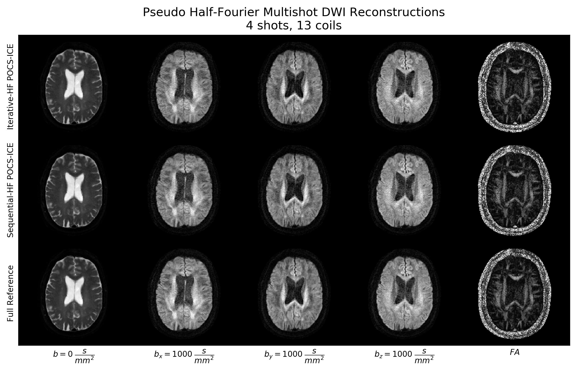
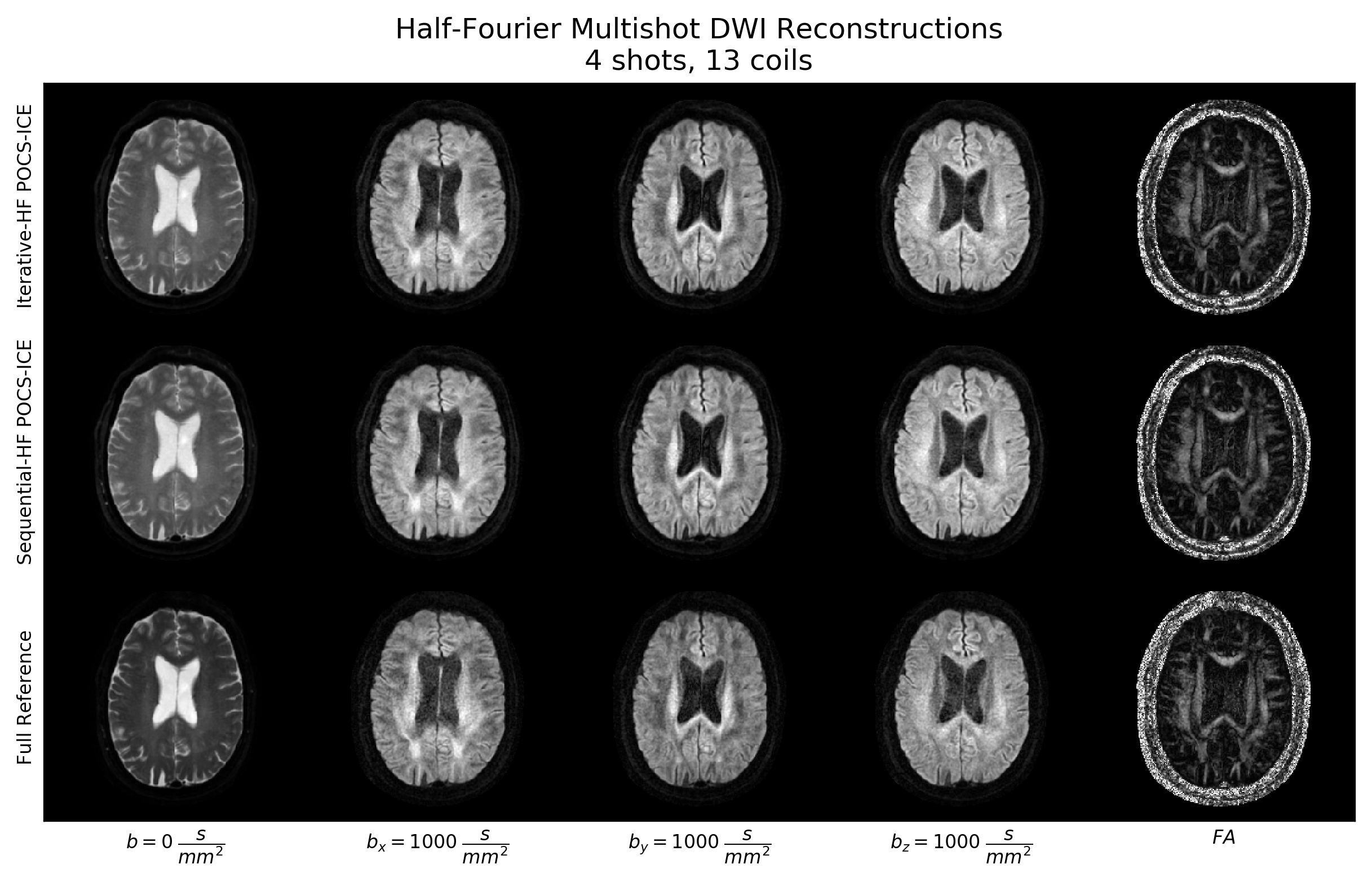
The algorithms were tested in pseudo half-Fourier simulations and in-vivo. In the simulations, full-Fourier multi-shot in-vivo brain data was acquired with $$$N_{shots}=\{3, 4, 6\}$$$ and 5 slices from 4 healthy subjects. This full data was reconstructed by POCS-ICE as reference and then reduced by 5/8th for HF testing. We decided to use in-vivo data for simulating under realistic phase conditions. For in-vivo evaluation, healthy brain data was acquired with $$$N_{shots}=4$$$ and $$$HF-factors=\{0.632, 1\}$$$ with echo times $$$T_E=\{65, 98\} \, ms$$$, respectively. Measurements were performed using a 13-channel head coil (3T Philips Ingenia), $$$b=\{0, 1000\} \, \dfrac{s}{mm^2}$$$ in three directions and $$$1 \times 1 \times 4 \, mm^3$$$ resolution. Informed consent was attained according to the rules of the institution.
Python 3.6.5 was used with a 2.7 GHz Intel Core i7 4-core CPU and 16 GB RAM. The algorithms were stopped when the residual error4 of subsequent iterations dropped below 10-3 or the iteration number exceeded 400. Motion-induced phase-estimation was disabled for $$$b=0 \, \dfrac{s}{mm^2}$$$.
Results
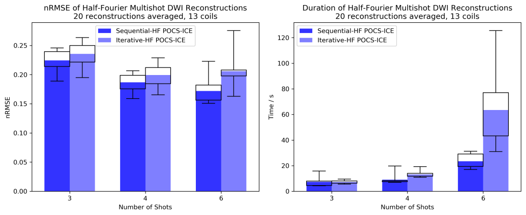
Normalized root-mean-square errors (nRMSE) and durations of the pseudo half-Fourier results are shown in Figure 2. Sequential-HF POCS-ICE has slightly lower nRMSE and is faster than Iterative-HF POCS-ICE.
For the full-Fourier in-vivo case, pseudo half-Fourier reconstructed diffusion images and fractional anisotropy (FA) maps are compared to the full POCS-ICE reference in Figure 3. Figure 4 analogously shows real half-Fourier reconstructions. Sequential-HF and Iterative-HF POCS-ICE results appear consistent with the reference and sharply resolve the anatomical brain structures.
Discussion and Conclusion
Both half-Fourier algorithms successfully recover in-vivo HF datasets with increased SNR and different contrast caused by the reduced echo time. The methods reveal ventricle microstructures for $$$b_0$$$ and $$$b_y$$$ that are not visible in the fully sampled reference. This is probably caused by the constrained phase within the diffusion-problematic ventricle and, moreover, emphasized by the higher SNR. Sequential-HF outperforms Iterative-HF POCS-ICE in computational speed. The nRMSE is comparable and remains nearly stable (Figure 2a) as the reference equally suffers from higher segmentation.
In conclusion, half-Fourier and multi-shot constraints were successfully combined in two novel algorithms achieving high-quality reconstructions in simulations and in-vivo paving the way for clinical adoption.
Acknowledgements
No acknowledgement found.References
1. Wu W and Miller KL. Image formation in diffusion MRI: A review of recent technical developments: Review of Image Formation in dMRI. JMRI. 2017;46(3):646–662.
2. Pruessmann KP, Weiger M, Scheidegger MB, Boesiger P. SENSE: sensitivity encoding for fast MRI. MRM. vol. 1999;42(5):952–962.
3. Pruessmann KP, Weiger M, Börnert P, Boesiger P. Advances in sensitivity encoding with arbitrary k‐space trajectories. MRM. 2001;46(4):638-651.
4. Guo H, Ma X, Zhang Z, Zhang B, Yuan C, Huang F. POCS‐enhanced inherent correction of motion‐induced phase errors (POCS‐ICE) for high‐resolution multishot diffusion MRI. MRM. 2016;75(1):169-180.
5. Chen N, Guidon A, Chang HC, and Song AW. A robust multi-shot scan strategy for high-resolution diffusion weighted MRI enabled by multiplexed sensitivity-encoding (MUSE). NeuroImage. 2013;72:41–47.
6. Liang ZP and Lauterbur PC. Principles of Magnetic Resonance Imaging: A Signal Processing Approach. IEEE Press. 2000.
7. Samsonov AA, Kholmovski EG, Parker DL, and Johnson CR. POCSENSE: POCS-based reconstruction for sensitivity encoded magnetic resonance imaging. MRM. 2004;52(6):1397–1406.
Figures