3338
Diffusion imaging of rat brain slices on a human clinical MRI scanner1Department of Electrical and Computer Engineering, University of Illinois at Urbana Champaign, Urbana, IL, United States, 2Beckman Institute for Advanced Science and Technology, Urbana, IL, United States, 3Department of Bioengineering, University of Illinois at Urbana Champaign, Urbana, IL, United States, 4Department of Cell and Developmental Biology, University of Illinois at Urbana Champaign, Urbana, IL, United States
Synopsis
We demonstrate
the feasibility of performing diffusion tensor imaging on a rat brain slice using a 3 T human clinical scanner and a rat coil. Brain slices provide an important platform for performing mechanistic studies in neuroscience. We show that sufficient SNR is available
for performing experiments to examine the diffusion properties of white matter through examining age-related differences in FA on 8-week-old and 1-year-old rats.
Introduction
The use of brain slices is an important tool to perform mechanistic studies in neuroscience. It enables the researcher to isolate aspects of physiological function and to explore the impact of specific signaling pathways, gene promoters, and inhibitors on the structure and function of particular brain regions. Many methods exist to reveal the microstructure of the tissue at the end of the brain slice experiment, such as Nissl staining; however, MRI provides an interesting opportunity to quantify dynamic changes during a longitudinal brain slice experiment. Diffusion tensor imaging (DTI) is frequently used to examine the integrity and myelination of white matter tracts as a function of aging, with decreases in fractional anisotropy (FA) with age in humans (Davis, 2009), but FA increases with age in rats (Yates, 2007). The use of DTI in brain slices enables mechanistic studies of aging, myelination, and the relationship to DTI measures, due to the lack of blood and subject movement as well as end-point histological staining. Given the increasing prevalence of clinical MRI scanners with improved gradient performance, in this work, we examine if DTI can be performed on a brain slice on a clinical MRI scanner.Methods
Scanning is done on a Siemens 3 T Prisma clinical scanner, with a transmit/receive Rapid Biomed volumetric rat coil. The length of the coil is 210 mm, with an inner diameter of 72mm. We used a hippocampal brain slice of a 4-month-old male LE BluGill rat and, to compare young and old rats, brain slices from an 8-week-old and a 1-year-old male rat. These brain slices are placed together in one dish. The thickness of the slices range from 2 mm-4 mm.
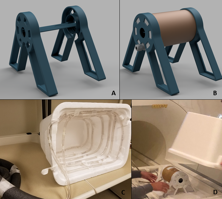
To enhance longevity of the brain slices, two important physical conditions are considered. The first is a constant 37°C temperature, which will also be crucial to any diffusion measurements taken. A Styrofoam box was used as a radiator, with plastic tubing lining the inside to heat the box. The second physical condition is stability inside the rat coil, to reduce motion artifacts and keep the brain slice from spilling out of the dish. A 3D printed holder kept the rat coil self-standing and securely positioned the dish at the coil’s isocenter (Figure 1).
A turbo spin-echo T2 weighted sequence was used to obtain a high-resolution structural scan, with a spatial resolution of 0.125mm, FOV of 40mm, matrix size 320, TR 2.5s and TE 181ms. For the diffusion tensor image, a spin-echo diffusion scan was used. Here, the spatial resolution was 0.352 mm in plane, FOV 45mm, matrix size of 128, TR 1.5s, and TE 70ms. The b value was 1000 s/mm2. Processing was then completed in FSL using the diffusion toolbox, FDT (Behrens, 2003).
To confirm that there was an adequate signal-to-noise ratio (SNR), the mean and standard deviation were taken over three averages, pixel by pixel, for each diffusion direction. A final SNR map was generated by averaging over the thirty directions.
Results
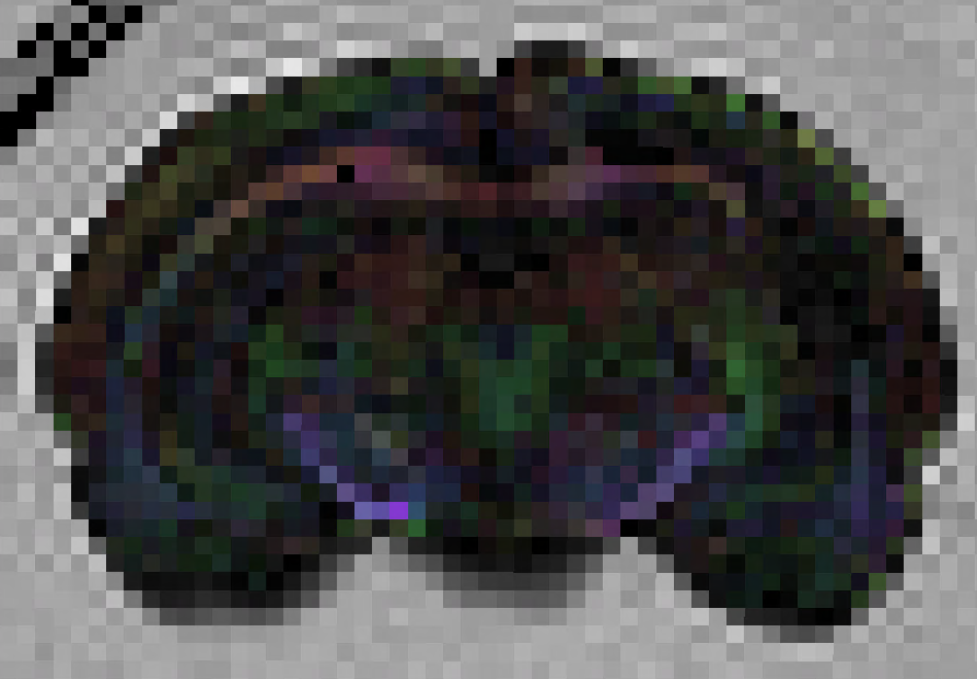
A color-coded FA map is given in Figure 2 indicating white matter structures such as the corpus callosum at the top of the image, and hippocampus. Average FA values were around 0.15.
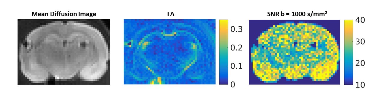
SNR maps (Figure 3) show that an adequate SNR is possible for diffusion tensor imaging. SNR values of around 40 are calculated in the majority of the brain tissue. As a comparison, the b=0 image has SNR values of 60.
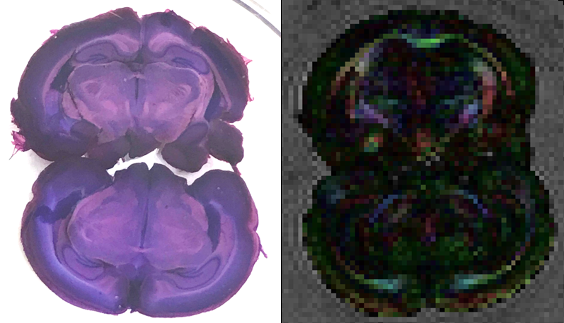
Differences in FA values between a young and old rat are shown in Figure 4. The 8-week-old rat displayed average values of 0.14 and the 1-year-old rat had higher FA values of 0.16. Averages were taken over white matter regions by thresholding FA values above 0.1 inside the brain.
Discussion
The SNR produced by the diffusion images was adequate to visualize properties of white matter in rat brain slices on a human clinical MRI scanner. Specific brain structures are visible in the FA images that agree with known rat brain atlases, such as the Waxholm Space Sprague Dawley atlas (Papp, 2014). We demonstrated sufficient signal and resolution for detecting differences in brain structure of aging rats, confirming that FA in rat brains increases with age, as previously noted (Yates, 2007).Conclusion
The results indicate that high spatial resolution at a small scale is achievable with a clinical MRI scanner. With the ability to successfully image rat brain slices with diffusion, many opportunities for mechanistic brain slice studies are available on commonly available clinical scanners.Acknowledgements
This work was supported by the NSF-NRT Grant 173525, Understanding the Brain: Training the Next Generation of Researchers in Engineering and Deciphering of Miniature Brain Machinery.References
[1] Behrens TEJ, Woolrich MW, Jenkinson M, Johansen-Berg H, Nunes RG, Clare S, Matthews PM, Brady JM, Smith SM. Characterization and propagation of uncertainty in diffusion-weighted MR imaging. Magn Reson Med. 2003; 50(5):1077-1088.
[2] Davis SW, Dennis NA, Buchler NG, White LE, Madden DJ, Cabeza R. Assessing the effects of age on long white matter tracts using diffusion tensor tractography. NeuroImage. 2009;46(2):530-41.
[3] Papp EA, Leergaard TB, Calabrese E, Johnson GA, Bjaalie JG. Waxholm Space atlas of the Sprague Dawley rat brain. NeuroImage. 2014;97:374-386.
[4] Yates MA, Juraska JM. Increases in size and myelination of the rat corpus callosum during adulthood are maintained into old age. Brain Res. 2007;1142:13–8.
Figures