3337
Development of High Quality T1w and DTI Templates of the Older Adult Brain in a Common Space1Biomedical Engineering, Illinois Institute of Technology, Chicago, IL, United States, 2Rush Alzheimer's Disease Center, Rush University, Chicago, IL, United States
Synopsis
The demand for a multimodal MRI atlas of the older adult brain is increasing as large amounts of data are generated in studies of aging. The purpose of this work was to develop high quality T1-weighted (T1w) and diffusion tensor imaging (DTI) templates of the older adult brain in the same space, to allow future multimodal analyses. This was successfully accomplished through a proposed iterative multimodal template construction strategy. The new templates allowed higher spatial normalization accuracy of T1w and DTI data from older adults compared to other available templates.
Introduction
Methods
Data and Pre-processing:
T1w and DTI data were collected on 202 non-demented older adults (50% male, 65.2-94.9 years of age) participating in the Rush Memory and Aging Project2, using a 3T MRI scanner. DTI data were processed (including undistortion) using TORTOISE3 and were aligned to the raw T1w images by applying the rigid and affine transformations derived from registering b=0 to T1w images.
T1w and DTI Template Construction Strategy:
In the proposed iterative template construction approach, each iteration includes two steps. In step 1, spatial normalization across the 202 participants is driven by T1w information, a temporary T1w template is generated, and the resulting transformations are also applied to the DTI data (Fig.1). In step 2, spatial matching is driven by DTI information, a temporary DTI template is generated, and the resulting transformations are also applied to the T1w data. Overall, both the T1w and DTI data experience the same transformations in both steps and all iterations (Fig.1). This approach ensures that each step maximizes the quality of the corresponding template (step 1 maximizes quality of T1w template, step 2 maximizes quality of DTI template), and spatial matching between the two templates increases for more iterations. Spatial normalization in step 1 is based on ANTs4, and in step 2 on DR-TAMAS5. The transformations from each step and iteration are combined to minimize interpolations.
Evaluation of T1w and DTI Templates at Different Iterations:
The quality of the T1w template generated at different iterations was assessed by means of the inter-subject standard deviation in gray matter of normalized T1w images used in the construction of the template. Quality of the DTI template was assessed by means of the average pair-wise Euclidean distance of tensors (DTED) as well as the coherence of primary eigenvectors (COH) across participants used in the construction of the template6. The spatial matching between T1w and DTI templates was assessed by means of the Spearman’s rank correlation between the white matter tissue probability map generated from the T1w template and the FA map of the DTI template7. Histograms of the above quantities were compared across iterations using a two-sample one-sided Kolmogorov-Smirnov test.
Comparison of the New Templates to Other Standardized Templates in Terms of Spatial Normalization Accuracy of External Data:
The final T1w and DTI templates were compared with other templates including older adults: {T1w templates: [MCALT v1.18, IXI-ANTs9, and IXI-Dartel10]; DTI template: [IXI aging DTI template V2.011]} in terms of the spatial normalization accuracy achieved when used as reference for normalization of data from older adults. The average pair-wise cross-correlation and standard deviation in gray matter of normalized T1w data were compared across T1w templates, and histograms of DTED and COH of normalized DTI data were compared across DTI templates.
Results and Discussion
Figure 2 demonstrates that the spatial matching of the T1w and DTI data used to construct the templates improves with more iterations (p<10-10), suggesting that the template quality also improves. Figure 3 shows that the spatial matching of the T1w and DTI templates also improves with more iterations (p<10-10). The improving quality and spatial matching of the templates with more iterations was also verified with visual inspection. Figures 4 and 5 demonstrate that the final T1w and DTI templates allow higher spatial normalization accuracy of T1w and DTI data from older adults compared to other available templates (p<10-10).Conclusion
The present work proposed a new iterative approach for construction of multimodal templates. The new approach generated high quality T1w and DTI templates of the older adult brain, in the same space. The new templates allowed higher spatial normalization accuracy of T1w and DTI data from older adults compared to other available templates.Acknowledgements
National Institute on Aging R01AG052200References
1. Weiner MW, Veitch DP, Aisen PS, et al. The Alzheimer's Disease Neuroimaging Initiative: a review of papers published since its inception. Alzheimers Dement. 2013;9(5):e111-94.
2. Bennett DA, et al. Curr Alzheimer Res. 2012;9:646-663.
3. Pierpaoli C, et al. TORTOISE: an integrated software package for processing of diffusion MRI data. Proc. Intl. Soc. Mag. Reson. Med. 2010; 18:1597.
4. Avants et al. ANTS: Open-Source Tools for Normalization And Neuroanatomy.
5. Irfanoglu M. O., Nayak, A., Jenkins, J., Hutchinson, E. B., Sadeghi, N., Thomas, C. P., et al. DR-TAMAS: diffeomorphic registration for tensor accurate alignment of anatomical structures. Neuroimage 2016;132:439–454.
6. Zhang S., et al. Enhanced ICBM diffusion tensor template of the human brain. Neuroimage. 2011;54(2):974-84.
7. Hsu Y. C., et al. NTU-DSI-122: A diffusion spectrum imaging template with high anatomical matching to the ICBM-152 space. Hum Brain Mapp 2015;36(9):3528–3541.
8. Christopher G., et al. The Mayo Clinic Adult Lifespan Template (MCALT): Better Quantification across the Lifespan. In Proc: Alzheimer's Association International Conference, 2017.
9. Avants, B., Tustison, N. ANTs/ANTsR Brain Templates. 2018;figshare. Fileset.
10. Ashburner, J. A fast diffeomorphic image registration algorithm. Neuroimage 2007;38:95–113.
11. Zhang, H., et al. A computational white matter atlas for aging with surface-based representation of fasciculi. Biomedical Image Registration. WBIR 2010. Lecture Notes in Computer Science, vol 6204.
12. Nooner K.B. et al. The NKI-Rockland Sample: A model for accelerating the pace of discovery science in psychiatry. Frontiers in neuroscience 2012;6:152.
Figures
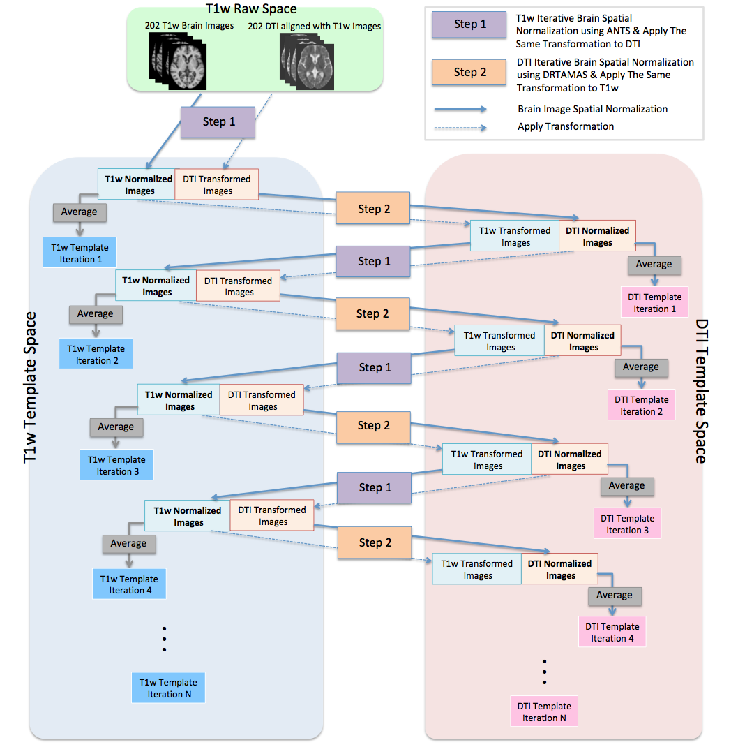
Figure 1.
Diagram of the proposed approach for the construction of T1w and DTI templates of the older adult brain.

Figure 2.
(a) Histograms of the relative number of gray matter voxels at different values of inter-subject standard deviation of normalized T1w images used in the construction of the template. (b) Histograms of the relative number of white matter voxels at different values of the average pair-wise Euclidean distance of tensors (DTED) across participants used in the construction of the DTI template. (c) Histograms of the relative number of white matter voxels at different values of the coherence of primary eigenvectors (COH) across participants used in the construction of the DTI template.
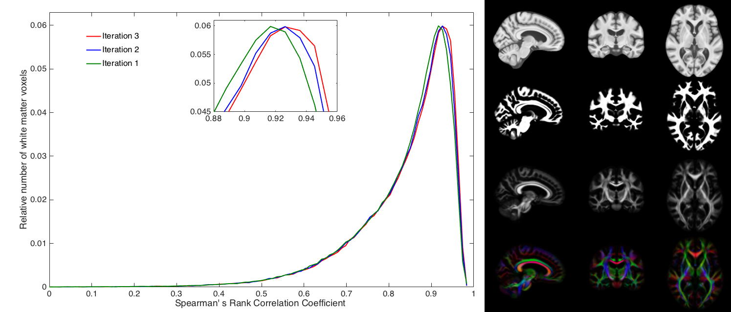
Figure 3.
Relative number of white matter voxels at different values of Spearman’s rank correlation between the white matter tissue probability map generated from the T1w template and FA map of the DTI template. The spatial matching of the T1w and DTI templates improves with more iterations. The right panel shows the final T1w template (1st row) and corresponding white matter tissue probability maps (2nd row), and the FA maps (3rd row) and anisotropy color maps (4th row) of the final DTI template.

Figure 4.
Evaluation of spatial normalization accuracy achieved for T1w data from 221 ADNI1 non-demented older adults (65-95 years) when using as reference the final T1w template, MCALT v1.1 (30-92 years), IXI-ANTs (20-90 years), IXI-Dartel (17-80 years). (a) Barplot of the average pair-wise cross-correlation of normalized T1w images. (b) Histograms of the relative number of gray matter voxels at different values of the inter-subject standard deviation of spatially normalized T1w images. The final T1w template allows higher spatial normalization accuracy for T1w data from older adults compared to other available templates.

Figure 5.
Evaluation of spatial normalization accuracy achieved for DTI data from 72 healthy older adults (65-85 years) from the Enhanced Nathan Kline Institute Rockland Sample12 when using as reference the final DTI template, or IXI Aging DTI template V2.0 (65-83 years). Histograms of the relative number of white matter voxels at different values of the average pair-wise Euclidean distance of tensors (DTED) as well as the coherence of primary eigenvectors (COH) across spatially normalized DTI data. The final DTI template allows higher spatial normalization accuracy for DTI data from older adults compared to the IXI Aging DTI template V2.0.