3333
Investigating the biodistribution of the antiinflammatory drug teriflunomide in vivo using 19F MRI1Berlin Ultrahigh Field Facility, Max Delbrück Center for Molecular Medicine, Berlin, Germany, 2Experimental and Clinical Research Center, a joint cooperation between the Charité Medical Faculty and the Max Delbrück Center for Molecular Medicine, Berlin, Germany
Synopsis
Teriflunomide is a trifluorinated drug indicated in the treatment of multiple sclerosis (MS). Using fluorine (19F) MR methods, the biodistribution of this anti-inflammatory drug could be examined in vivo to guide pharmacological studies and dosage adjustments en route to individualized therapy. In this study, we administered teriflunomide to healthy rats and an animal model of MS. We could detect teriflunomide non-invasively in various tissues in vivo, during the disease course and ex vivo.
Introduction
A high variability in in vivo drug activity makes the treatment of multiple sclerosis (MS) a challenge and limits the likelihood for predicting treatment outcome1. Since one third of all approved drugs are fluorinated, these could be non-invasively detected and quantified during pathologies by 19F MR techniques2-4. The ultimate goal is to guide pharmacokinetic studies and enable precise adjustments of the drug dose. In this study we assessed the distribution of the anti-inflammatory compound teriflunomide5 at its target organ the brain in vivo at different stages of the disease in the experimental autoimmune encephalomyelitis (EAE) model.Methods
Female Dark Agouti rats and SJL/J mice were used for studying the distribution of teriflunomide in vivo. EAE was induced by subcutaneously immunizing female SJL/J (3 months) with the CNS proteolipid protein (PLP139–151, 250µg), emulsified with M.Tuberculosis H37RA (800µg) in 100µl Complete Freund's Adjuvant6. Pertussis Toxin (1.25ng/µl in 200µl PBS) was administered intraperitoneally on days 0 and 2. A neurologic scoring was performed daily to assess the EAE symptoms (righting reflex weakness 0.5, tail paresis 0.5, tail paralysis 1, unilateral hindlimb paresis 0.5, bilateral hindlimb paralysis 1, unilateral forelimb paresis 0.75, bilateral forelimb paralysis 1.5). Teriflunomide was prepared in carboxymethylcellulose (CMC)7,8 and animals were treated daily with 30mg/kg teriflunomide by oral gavage, using a blunt-end needle9. MR experiments were performed on a Bruker Biospec 9.4T MR Scanner (Bruker, Ettlingen, Germany), using a room temperature 19F/1H RF-coil10 and a cryogenically-cooled 19F RF-coil (CRP)11. FLASH and 3D-RARE were used for anatomical imaging. Brain lesions in EAE mice were detected using MDEFT (TR/TE/TI 2600/3.9/950ms, FOV 30.2x12.8x9mm, Matrix 256x170x18, TA=3m 7s) before and after administration of gadolinium (Magnevist) as contrast agent. Global single-pulse 19F MRS (TR=1000ms, TA=17min) was used to detect the 19F signal. After in vivo studies, animals were killed under narcosis and tissues were fixed, for further ex vivo studies.Results
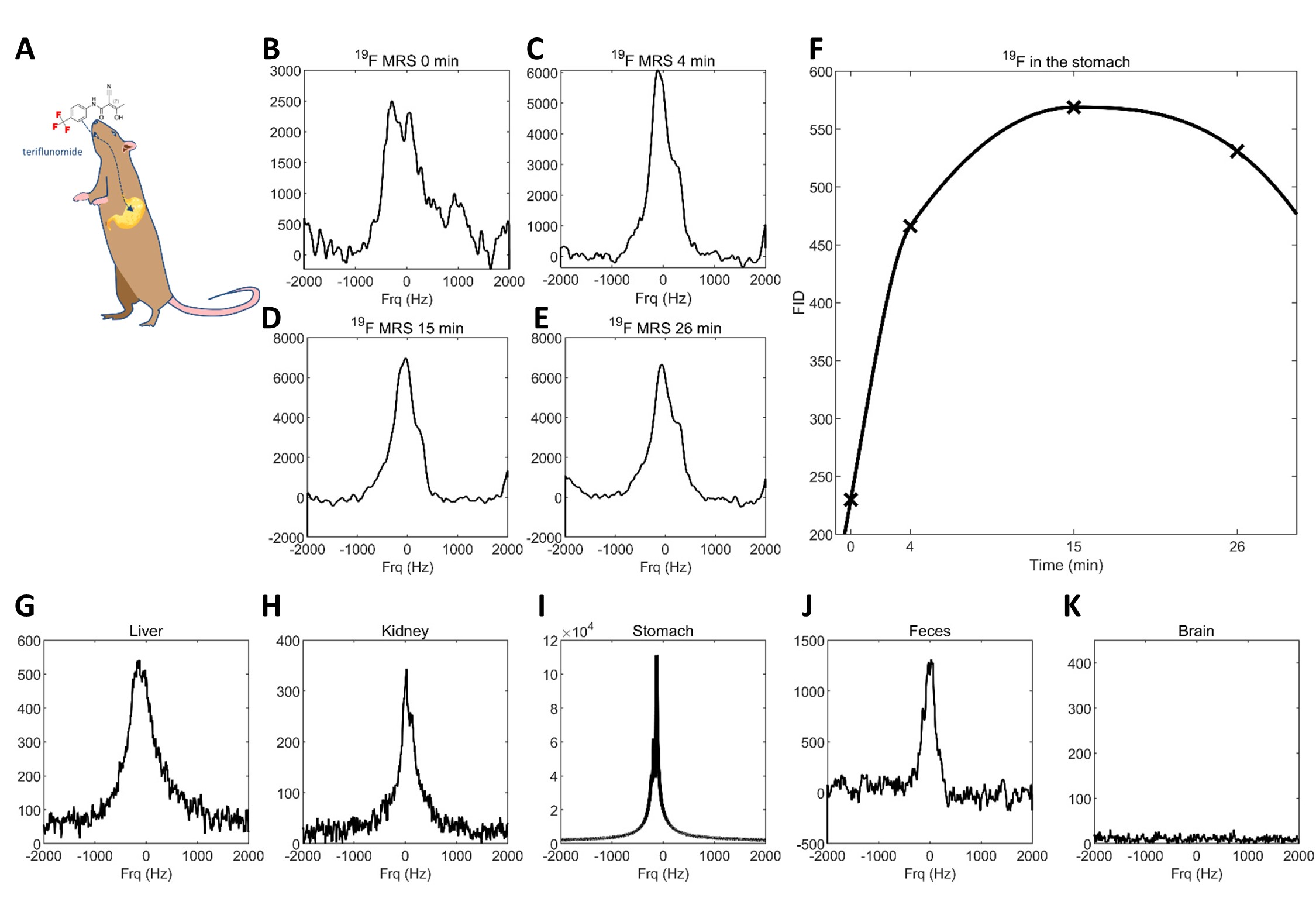
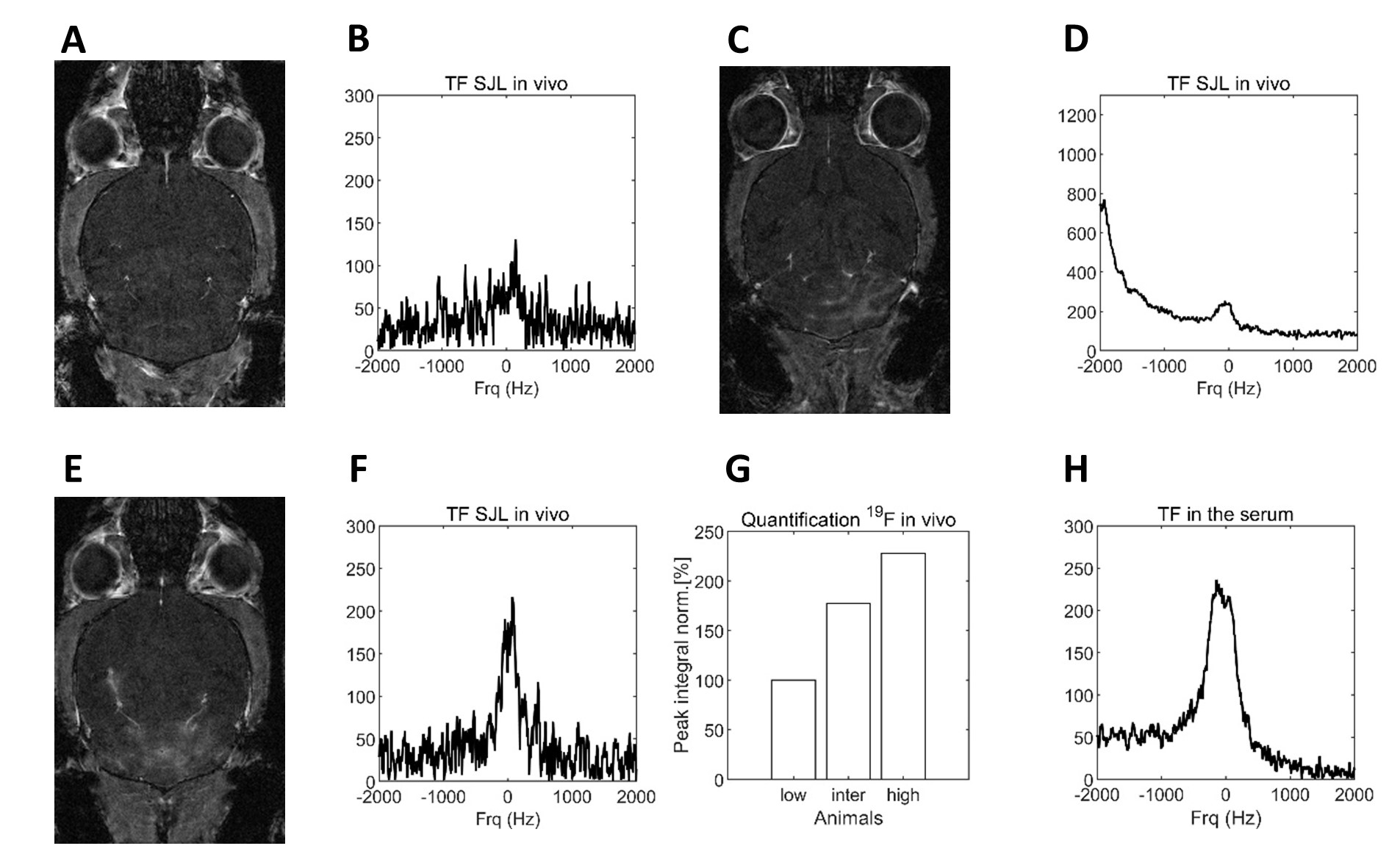
Teriflunomide was detected in healthy Dark Agouti rats in vivo by 19F MRS. TF was administered orally via gastric intubation (Fig.1A) during MR acquisitions. Repetitive 19F MRS (Fig.1B-E) was acquired for over half an hour (Fig.1F). During the first 15min, the signal intensity increased, possibly due to the chemical environment (lower pH) in the stomach that renders TF soluble in CMC. The 19F signal decreased over time suggesting an absorption of TF from the small intestine into the blood stream. The distribution of TF was studied ex vivo by 19F MRS using the 19F CRP (Fig.1G-K). TF was detected in the liver (G), kidney (H) stomach content (I) and feces (J), but not in the brain (K). 19F MR signals were also studied in EAE mice treated with TF (30mg/kg) daily over a period of 10 days following disease induction. First, the extent of CNS pathology was determined by studying inflammatory lesion load following gadolinium contrast application (Fig.2A+D+E). A 19F MR signal could be detected in the head region of all EAE mice scanned in vivo with the 19F CRP even in EAE mice that presented with no neurological score and no contrast-enhanced brain lesions (Fig.2A). For the latter the 19F MR signal was small (Fig.2B). In an EAE mouse with a neurological score of 0 but presence of gadolinium-enhancing lesions (Fig.2C) an intermediate 19F MR peak was detected (Fig.2D). In the EAE mouse with the highest score (1.25), that also presented with gadolinium-enhanced brain lesions (Fig.2E) a substantial 19F peak could be detected by 19F-MRS (Fig.2F). The signal amount was quantified using the integral of the spectra (Fig.2G). TF was also detected in the serum of these animals ex vivo (Fig.2H).Discussion
The detection of teriflunomide in the stomach of healthy rats demonstrates the first pharmacokinetic step following oral administration. We followed the 19F signal in the stomach over time and detected the drug in various tissues ex vivo as a first step towards studying the distribution in vivo. Our in vivo experiments in EAE mice brain might indicate a connection between 19F MR signal and disease severity, which might be a result of blood-brain-barrier leakage leading to differences in drug distribution within the CNS. Such changes are expected to influence the efficacy, with regard to symptoms and disease course. Further experiments are required to confirm this observation.Conclusions
This study showed the first detection of TF using 19F MR. Following a thorough investigation of the MR properties12, the present results are the next step towards advancing 19F MR methods for detecting TF therapy in vivo. Tracking 19F compounds non-invasively could provide invaluable insights into drug distribution especially during pathology and aims providing patient tailored therapy monitoring.Acknowledgements
This work was funded by Sanofi-Aventis. This work was supported by funding from the Germany Research Council (DFG WA2804).References
1 Grossman, I. et al. Pharmacogenomics strategies to optimize treatments for multiple sclerosis: Insights from clinical research. Progress in neurobiology 152, 114-130, doi:10.1016/j.pneurobio.2016.02.001 (2017).
2 Müller, K., Faeh, C. & Diederich, F. Fluorine in pharmaceuticals: looking beyond intuition. Science 317, 1881-1886 (2007).
3 Niendorf, T., Ji, Y. & Waiczies, S. in Fluorine Magnetic Resonance Imaging (ed E. T. Ahrens, Flögel U.) 311-344 (Pan Stanford Publishing, 2016).
4 Reid, D. G. & Murphy, P. S. Fluorine magnetic resonance in vivo: a powerful tool in the study of drug distribution and metabolism. Drug discovery today 13, 473-480, doi:10.1016/j.drudis.2007.12.011 (2008).
5 Desmoulin, F., Gilard, V., Malet-Martino, M. & Martino, R. Metabolism of capecitabine, an oral fluorouracil prodrug: (19)F NMR studies in animal models and human urine. Drug metabolism and disposition: the biological fate of chemicals 30, 1221-1229 (2002).
6 Lepore, S. et al. Enlargement of cerebral ventricles as an early indicator of encephalomyelitis. PloS one 8, e72841, doi:10.1371/journal.pone.0072841 (2013).
7 Ringheim, G. E. et al. Teriflunomide attenuates immunopathological changes in the dark agouti rat model of experimental autoimmune encephalomyelitis. Frontiers in neurology 4, 169, doi:10.3389/fneur.2013.00169 (2013).
8 Iglesias-Bregna, D. et al. Effects of prophylactic and therapeutic teriflunomide in transcranial magnetic stimulation-induced motor-evoked potentials in the dark agouti rat model of experimental autoimmune encephalomyelitis. J Pharmacol Exp Ther 347, 203-211 (2013).
9 Merrill, J. E. et al. Teriflunomide reduces behavioral, electrophysiological, and histopathological deficits in the Dark Agouti rat model of experimental autoimmune encephalomyelitis. J Neurol 256, 89-103 (2009).
10 Waiczies, H. et al. Visualizing brain inflammation with a shingled-leg radio-frequency head probe for 19F/1H MRI. Scientific reports 3, 1280, doi:10.1038/srep01280 (2013).
11 Waiczies, S. et al. Enhanced Fluorine-19 MRI Sensitivity using a Cryogenic Radiofrequency Probe: Technical Developments and Ex Vivo Demonstration in a Mouse Model of Neuroinflammation. Scientific reports 7, 9808, doi:10.1038/s41598-017-09622-2 (2017).
12 Prinz, C. et al. 19F MR characterization of teriflunomide, a fluorinated drug indicated in Multiple Sclerosis. Proc ISMRM (2018).
Figures