3330
Regional morphometric abnormalities and clinical relevance in Wilson's disease1Department of Radiology, The First Affiliated Hospital of Xiamen University, Xiamen, China, 2Department of Radiology, The First Affiliated Hospital of Sun Yat-sen University, Guangzhou, China, 3Department of Electrical and Electronic Engineering, Southern University of Science and Technology, Shenzhen, China, 4Southern University of Science and Technology, Shenzhen, China
Synopsis
In this study, coarse-to-fine evaluations were creatively made in terms of both global volume and local shape to identify Wilson’s disease (WD)-related morphometric abnormalities of eight structures of interest (caudate, putamen, globus pallidus, thalamus, amygdala, hippocampus, red nucleus and substantia nigra). Our results revealed that significant volume reductions and region-specific surface atrophy were detected in all structures of interest except the bilateral hippocampus in patients with WD relative to HC subjects, and the putamen had the strongest global and local atrophy and the amygdala was least affected. These morphometric abnormalities may serve as useful imaging biomarkers for WD.
INTRODUCTION
Wilson’s disease is an inherited autosomal recessive disorder of copper metabolism leading to copper accumulation in hepatocytes and in extrahepatic organs such as the brain and the cornea. 1, 2 The copper accumulation in the brain can induce changes such as myelin degeneration, gliosis and profound neuronal loss. 3, 4 Among all brain structures, the gray matter in the brain stem nuclei and the basal ganglia are most affected. 5-6 In this study, we aimed to detect and quantify regional morphometric abnormalities, in terms of both volume and shape, in patients with WD.METHODS
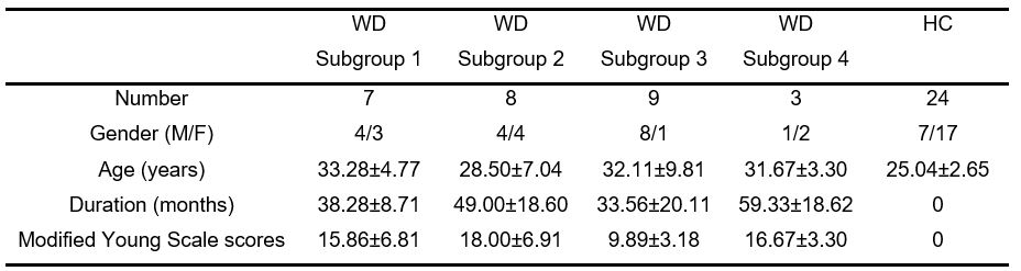
Twenty-seven WD patients and twenty-four healthy controls (HCs) were enrolled. Specific brain structures, including the bilateral caudate, putamen, globus pallidus, thalamus, amygdala, hippocampus, red nucleus and substantia nigra were automatically extracted from each participant’s T1-weighted image. Volume abnormalities and correlations to the modified Young scale were investigated. Furthermore, vertex-based shape analysis was performed to explore region-specific morphometric abnormalities.RESULTS
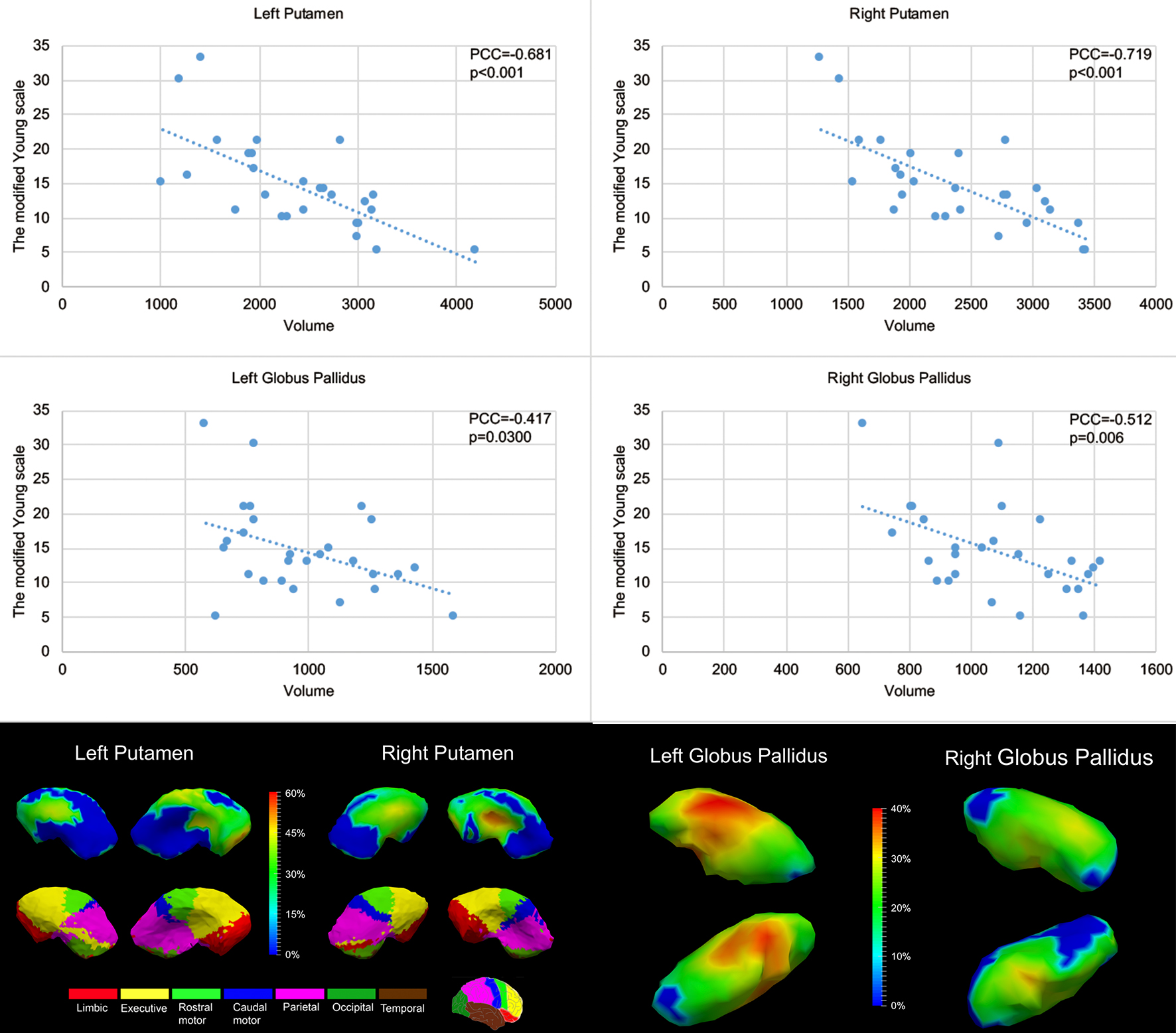
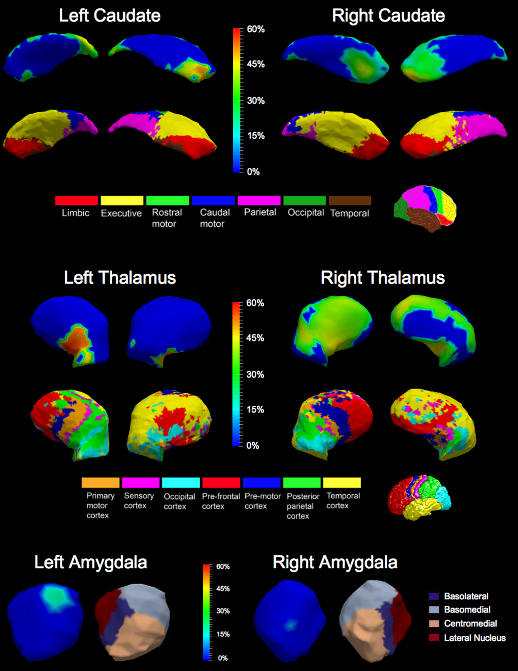
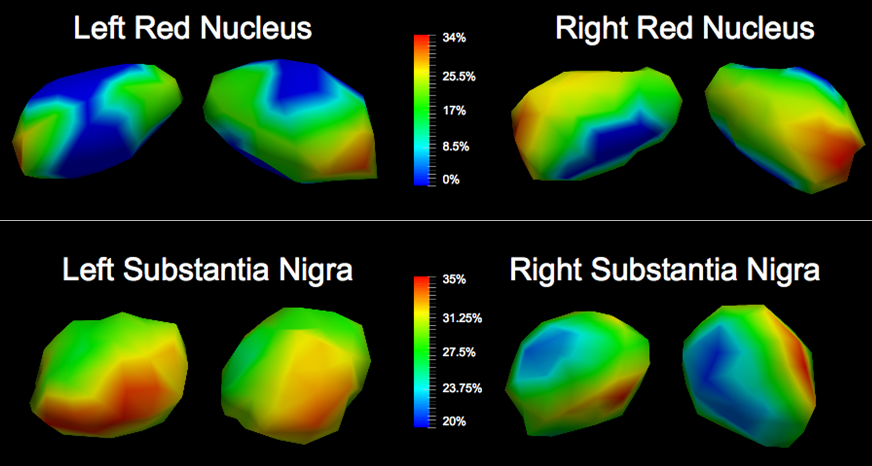
Significant global volume atrophy and local shape abnormalities were detected and quantified in the bilateral caudate, putamen, globus pallidus, thalamus, amygdala, red nucleus and substantia nigra in WD. Morphometric abnormalities of the caudate, putamen and thalamus were strong whereas those of the globus pallidus, amygdala, red nucleus and substantia nigra were milder. No hippocampal abnormalities were observed. The modified Young scale was found to correlate significantly with the volumes of the bilateral putamen and globus pallidus. Shape analysis revealed subregion-specific atrophy of the bilateral caudate and putamen, being concentrated on the subregions projecting to the limbic and executive cortices. Significant region-specific atrophy was also detected at the bilateral thalamic subregions projecting to the primary motor, sensory and premotor cortices.DISCUSSION
Most WD patients have neurological symptoms, including involuntary movements, speech disturbances, dysphagia, autonomic dysfunction as well as gait and balance disturbances. The basal ganglia, red nucleus and substantia nigra play important roles in a wide range of sensorimotor, cognitive and behavioral processes. 7 The thalamus, traditionally characterized as the central sensory and motor relay station of the brain, plays a key role in sleep, arousal, primary sensory processing and cognitive processes.8, 9 As such, atrophy of these structures may be closely linked to the neurological symptoms of WD. The subregion analysis results suggest differential contributions of distinct subregions to the clinical manifestations of WD, which is highly useful in understanding the neuropathology of WD. The executive cortex located in the frontal lobe is associated with executive processes. 10 The parietal cortex serves as a vital part in receiving and transforming sensory information into motor signals for sensory guidance of movements. 11 So, the atrophy of these subregional structures may be consistent with certain neurological symptoms of WD. Moreover, significant negative correlations were also observed between the modified Young scale and the volumes of the bilateral putamen and globus pallidus in the WD patients, indicating a potential relationship between the severity of clinical manifestations in WD and those structures’ volumes.CONCLUSION
This study demonstrated significant morphometric abnormalities of specific structures of interest in patients with WD, both globally and locally. These morphometric abnormalities may serve as useful imaging biomarkers for WD and further deepen our understanding of the neuropathology of WD.Acknowledgements
No acknowledgement found.References
1 ALA A, WALKER A P, ASHKAN K, et al. Wilson's disease[J]. Lancet, 2007,369(9559):397-408.
2 MEENAKSHI-SUNDARAM S, MAHADEVAN A, TALY A B, et al. Wilson's disease: A clinico-neuropathological autopsy study[J]. Journal of Clinical Neuroscience, 2008,15:409-417.
3 Mikol J, Vital C, Wassef M, et al. Extensive cortico-subcortical lesions in Wilson’s disease: clinico-pathological study of two cases. Acta Neuropathol. 2005;110(5):451-458.
4 Algin O, Taskapilioglu O, Hakyemez B, et al. Structural and neurochemical evaluation of the brain and pons in patients with Wilson’s disease. Jpn J Radiol. 2010;28(9):663-671.
5 Kuwert T, Hefter H, Scholz D, et al. Regional cerebral glucose consumption measured by positron emission tomography in patients with Wilson’s disease. Eur J Nucl Med. 1992;19(2):96-101.
6 Schlaug G, Hefter H, Engelbrecht V, et al. Neurological impairment and recovery in Wilson’s disease: evidence from PET and MRI. J Neurol Sci. 1996;136(1-2):129-139.
7 Groenewegen HJ. The Basal Ganglia and Motor Control. Neural Plast. 2003;10(1-2):107-120.
8 Adams RD, Victor M, Ropper AH, Daroff RB. Principles of neurology. 1997.
9 Van Der Werf YD, Tisserand DJ, Visser PJ, et al. Thalamic volume predicts performance on tests of cognitive speed and decreases in healthy aging. Cogn Brain Res. 2001;11(3):377-385.
10 Tziortzi AC, Haber substantia nigra, Searle GE, et al. Connectivity-Based Functional Analysis of Dopamine Release in the Striatum Using Diffusion-Weighted MRI and Positron Emission Tomography. Cereb Cortex. 2014;24(5):1165-1177.
11 Fogassi L, Luppino G. Motor functions of the parietal lobe. Curr Opin Neurobiol. 2005;15(6):626-631.
Figures