3325
Longitudinal brain volume changes in pre-menopausal breast cancer patients treated with chemotherapy1Imaging and Pathology, KU Leuven, Leuven, Belgium, 2Oncology, KU Leuven, Leuven, Belgium, 3Neurosciences, KU Leuven, Leuven, Belgium, 4Psychiatry, KU Leuven, Leuven, Belgium, 5Surgical Oncology, KU Leuven, Leuven, Belgium, 6Radiology, KU Leuven, Leuven, Belgium, 7Gynaecology and Obstetrics, KU Leuven, Leuven, Belgium, 8General Medical Oncology, KU Leuven, Leuven, Belgium, 9Center for Gynaecologic Oncology, Anthoni van Leeuwenhoek, Netherlands Cancer Insitute, Academical Medical Center, Amsterdam, Netherlands
Synopsis
This longitudinal study investigates possible recovery of volumetric brain changes in pre-menopausal patients three years after being treated for early-stage breast cancer. While initial widespread white matter volume increase was previously observed, recovery is seen three years after treatment in the same group of young women treated with chemotherapy. Patients with breast cancer show widespread gray matter volume decrease, observed both in patients treated and not treated with cytotoxic agents. Further studies are necessary to unravel possible acute volumetric changes, possibly neuro-inflammatory mechanisms, in this population as a cause for these findings.
Introduction
An increasing number of studies report chemotherapy-induced cognitive impairment and corresponding neuroimaging changes in cancer patients1–3. In a previous longitudinal deformation-based morphometry (DBM) study we reported white matter (WM) volume increase approximately six months after treatment with chemotherapy (t2) compared to baseline (t1), only significantly present in pre-menopausal patients4. This was in concordance with previous pre-clinical work, showing chemotherapy administration to cause frontal brain volume enlargement in a mouse model of breast cancer5. The current study investigates whether the previously observed volumetric brain changes are still present 3 years after the end of chemotherapy (t3).Methods
The initial population consisted of 180 women who were previously studied at t1 and t24,6; 72 women with early-stage breast cancer who underwent adjuvant chemotherapy (C+), 49 patients with breast cancer who did not receive chemotherapy (C-) and 59 healthy controls (HC). In this study, a subset of 51 young women were re-assessed three years after treatment or at matched interval for the HC (t3); 26 C+, 13 C- and 11 HC. All patients of this subset were pre-menopausal at baseline. T1-weighted 3D-TFE images (TR/TE/TI = 9.6/4.6/900ms, FOV 250x250x218mm, voxel size 0.98x0.98x1.20mm) were acquired from all participants on a Philips Intera 3.0T scanner with an 8-channel head coil. Volumetric brain changes were assessed with deformation-based morphometry. All images were preprocessed using ANTs N4-biasfield correction7 and the longitudinal pipeline of the CAT12 SPM12 (v6906) toolbox8. Probability maps of the Jacobian of the deformation matrices were calculated. The interaction effect between time of scan (t1 vs. t3) and treatment (C+, C- and HC) was analyzed using a GLM with Beck Depression Index (BDI) and scanner maintenance as covariates. All reported p-values were FWE-corrected on cluster level at α=0.05.Results
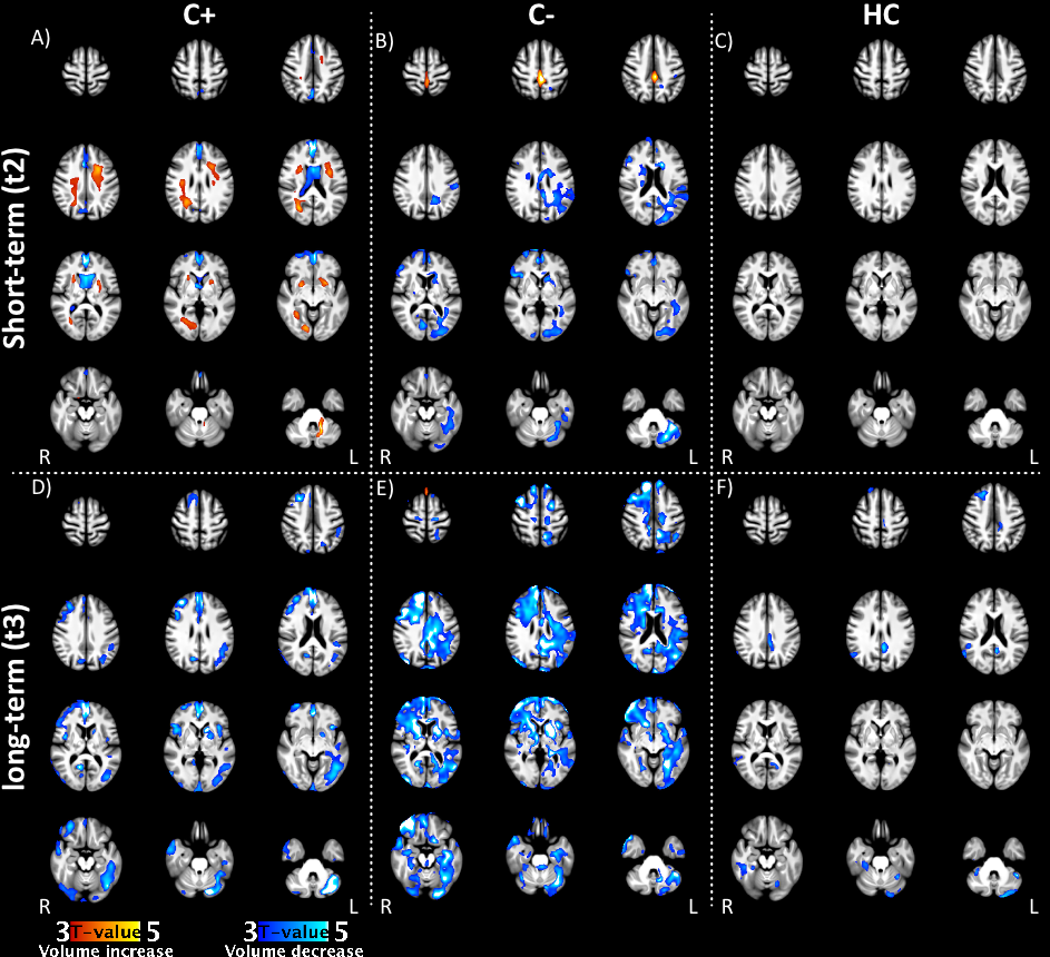
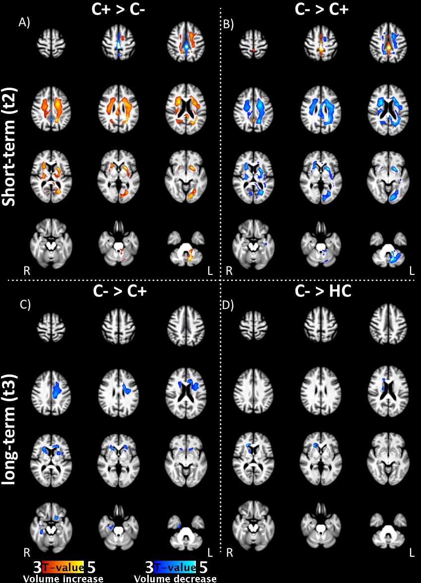
Initial white matter volume increase observed five to six months after chemotherapy4 (Figure 1:A,B,C) appeared to recover in a group of young breast cancer survivors three years after treatment (Figure 1:C). In contrast, decreased grey matter (GM) volume was seen in all groups three years later (Figure 1: D -F), with the most prominent decrease seen in C- patients (Figure 2: C-D). More specifically, C+ patients showed a diffuse pattern of GM volume decrease, most notably in the left cerebellar hemisphere, the medial prefrontal cortex, the left caudate head, the inferior frontal gyri and the right middle frontal gyrus; for the C- group a widespread decrease in GM and WM was found, combined with a volume increase in the frontal dorsal interhemispheric fissure; the HC group showed volume decreases in the cerebellum, the left posterior cingulate cortex and the right superior frontal gyrus.Discussion
The widespread GM volume decrease observed three years after chemotherapy is in line with earlier findings from voxel-based morphometry analyses9,10. Recovery from the observed WM volume increase in a group of pre-menopausal breast cancer patients is in agreement with previous diffusion tensor imaging findings in the same sample, which showed recovery of fractional anisotropy values in the WM in the same time-frame6. A potential mechanism underlying such microstructural changes could be edema formation, as an acute chemotherapy-induced neuro-inflammatory mechanism11, resolving over time (t3). Additionally, this study showed limited long-term GM volume decrease in healthy controls, which can be related to normal aging12. More pronounced GM decrease was observed in C- patients, raising the hypothesis of cancer therapy13 and/or hormonal changes14,15 resulting in accelerated ageing. This observed volume decrease was less pronounced in the C+ group compared to the C- group three years after chemotherapy. An initial volume increase4, possibly induced by neuro-inflammation, could partially mask the GM volume reduction.Conclusion
The results of this study confirm earlier findings of decreased GM volume observed after cancer treatment. Furthermore, we demonstrated that chemotherapy-induced WM volume expansion is no longer present three years after treatment. Future investigation is required to study a possible relation with neuro-inflammatory processes, as well as the neurocognitive impact of these findings.Acknowledgements
No acknowledgement found.References
1. McDonald BC, Saykin AJ. Alterations in brain structure related to breast cancer and its treatment: chemotherapy and other considerations. Brain Imaging Behav. 2013;7(4):374-387.
2. de Ruiter MB, Reneman L, Boogerd W, et al. Late effects of high-dose adjuvant chemotherapy on white and gray matter in breast cancer survivors: Converging results from multimodal magnetic resonance imaging. Hum Brain Mapp. 2012;33(12):2971-2983.
3. Li M, Caeyenberghs K. Longitudinal assessment of chemotherapy-induced changes in brain and cognitive functioning: A systematic review. Neurosci Biobehav Rev. May 2018.
4. Blommaert J, Amant F, Peeters R, et al. Longitudinal assessment of morphometric brain changes after chemotherapy in pre- and post-menopausal breast cancer patients. In: ESMRMB Conference Barcelona. ; 2017. (article currently under review)
5. Winocur G, Berman H, Nguyen M, et al. Neurobiological Mechanisms of Chemotherapy-induced Cognitive Impairment in a Transgenic Model of Breast Cancer. Neuroscience. 2018;369:51-65.
6. Billiet T, Emsell L, Vandenbulcke M, et al. Recovery from chemotherapy-induced white matter changes in young breast cancer survivors? Brain Imaging Behav. January 2017.
7. Tustison NJ, Avants BB, Cook PA, et al. N4ITK: Improved N3 Bias Correction. IEEE Trans Med Imaging. 2010;29(6):1310-1320.
8. Gaser C, Dahnke R. CAT - A Computational Anatomy Toolbox for the Analysis of Structural MRI Data. In: HBM Conference 2016. ; 2016.
9. Conroy SK, McDonald BC, Smith DJ, et al. Alterations in brain structure and function in breast cancer survivors: effect of post-chemotherapy interval and relation to oxidative DNA damage. Breast Cancer Res Treat. 2013;137(2):493-502.
10. Koppelmans V, de Ruiter MB, van der Lijn F, et al. Global and focal brain volume in long-term breast cancer survivors exposed to adjuvant chemotherapy. Breast Cancer Res Treat. 2012;132(3):1099-1106.
11. Ahles TA, Saykin AJ. Candidate mechanisms for chemotherapy-induced cognitive changes. Nat Rev Cancer. 2007;7(3):192-201.
12. Good CD, Johnsrude IS, Ashburner J, et al. A voxel-based morphometric study of ageing in 465 normal adult human brains. Neuroimage. 2001;14(1 Pt 1):21-36.
13. Ahles TA, Root JC. Cognitive Effects of Cancer and Cancer Treatments. Annu Rev Clin Psychol. 2018;14(1):annurev-clinpsy-050817-084903.
14. Boele FW, Schilder CMT, de Roode M-L, Deijen JB, Schagen SB. Cognitive functioning during long-term tamoxifen treatment in postmenopausal women with breast cancer. Menopause. 2015;22(1):17-25.
15. Seliktar N, Polek C, Brooks A, Hardie T. Cognition in breast cancer survivors: hormones versus depression. Psychooncology. 2015;24(4):402-407.
Figures