3322
Quantitative Data Driven Voxel-Wise Simulator (QVS): Application to 3D MP-RAGE Optimization for Harmonization of Multi-Centre Brain MRI Studies1Department of Brain Repair and Rehabilitation,Institute of Neurology, University College London, London, United Kingdom, 2Lysholm Department of Neuroradiology, National Hospital for Neurology and Neurosurgery, UCLH NHS Trust, London, United Kingdom, 3Wellcome Centre for Human Neuroimaging, UCL Queen Square Institute of Neurology, London, United Kingdom, 4Leonard Wolfson Experimental Neurology Centre, UCL Queen Square Institute of Neurology, London, United Kingdom
Synopsis
Typical MRI simulators are either based on the signal equations or on the solution to the Bloch equations for each voxel, with appropriate image encoding incorporated. In this work, a novel modular approach to simulate MR sequences, termed quantitative voxel-wise simulator (QVS), is proposed. This simulator employs quantitative parametric maps as inputs, models the evolved MR signal as a multi-dimensional filter output and reconstructs the image by using a voxel-by-voxel modulated k-space approach. As a proof-of-concept, 3D MP-RAGE sequence is simulated and the simulated images are compared with in-vivo data. QVS is designed for multi-centre brain MRI harmonization.
Introduction
An MRI simulator is a software tool that generates realistic multidimensional images according to the physical principles underlying the imaging process, using the known MR tissue properties. Executing MRI sequences with optimum performance requires appropriate and careful setting of numerous sequence parameters and MR simulators are a convenient tool for optimized parameter selection. Here we present a novel simulator that generates realistic human brain images for the 3D MP-RAGE sequence, which is widely used for morphological neuroimaging studies. The novel aspect of this simulator, termed Quantitative Voxel-Wise Simulator (QVS), is that it employs quantitative parametric maps as the input for solving the Bloch equations and a voxel-wise approach for image reconstruction.Methods
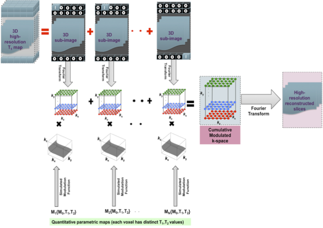
In the proposed simulator, the object is modeled as a discrete set of voxels, with each voxel characterized by a magnetization vector with tissue and sequence specific parameters. The tissue specific parameters include M0, T1, and T2, and the sequence specific parameters include flip angle (FA), repetition time (TR), etc. Based on this object model, separate electromagnetic field distributions i.e., main magnetic field inhomogenities (∆B0), RF transmit field inhomogenities (B1+) and RF receive field inhomogenities (B1-) corresponding to each voxel can be integrated in the voxel-wise simulation to account for scan dependent effects introduced by the MR image acquisition process. These distributions and the aforementioned tissue specific parameters were measured in volunteers using a multi-parameter mapping (MPM) acquisition.1 Discrete-event Bloch equation simulation was performed based on the MPM data i.e., quantitative T1 map, quantitative proton density (PD) map and the B1+ field map. In the case of 3D MP-RAGE, this magnetization simulation module generated a 3D filter or modulation function2 in response to the excitation and gradients applied during multiple TR. This procedure is depicted in Fig.1. Such filters were calculated for each voxel of the 3D volume. The following approach was taken in order to reconstruct the synthetic MP-RAGE images. Each voxel was individually Fourier transformed to obtain a synthetic k-space per voxel as shown in Fig. 1. Each voxel’s synthetic k-space was then multiplied by its respective filter function, calculated from the Bloch equation simulations. All filtered synthetic k-spaces were added together, and an inverse Fourier transform was applied to reconstruct the full simulated structural image. In order to compare the simulated output with the real data, ADNI33 MP-RAGE images were acquired on a 3T Siemens Prisma using a 64 channel head/neck array coil.Results
The quantitative R1-weighted map, which was obtained by using the MPM data processing toolbox,4 is shown in Fig. 2. The T1 values (T1=1/R1) and the PD values, calculated for each voxel, were used as input to the sequence (MP-RAGE) specific Bloch equation simulator. B1+ maps were used as input to the simulator to generate the effect of spatially varying flip angle and the bias field expected from the array coil on the simulated filter function. All sequence parameters for simulation were set according to the ADNI3 MP-RAGE parameters, excluding the parallel imaging factor.3 A representative slice of the simulated 3D MP-RAGE dataset is shown in Fig. 3. The respective slice from the acquired ADNI3 MP-RAGE image is shown in Fig. 4.Discussion and Conclusion
The QVS is a novel approach to simulate realistic structural MR images by using quantitative parametric maps as inputs. The simulated MP-RAGE images depict the expected grey-white matter contrast and match well with images acquired in the same volunteers with the same protocol. Both the simulated and acquired images are affected by blurring due to the intrinsic point-spread-function (PSF) of the MP-RAGE acquisition scheme. The MP-RAGE data was obtained with the ‘pre-scan normalise’ filter enabled to remove receive coil sensitivity effects; no filter was applied to the simulated data. Further investigation of the differences between the simulated and acquired images is underway. This simulator architecture has flexibility for incorporating spatially varying noise in the simulated MR images, which can vary with scanner hardware. Moreover, it is a versatile tool to analyze the performance of acquisition acceleration approaches such as parallel imaging or compressed sensing, for any arbitrary k-space undersampling patterns.Acknowledgements
This research is funded by the Alzheimer's Research United Kingdom (ARUK).References
1. Weiskopf Nikolaus, Suckling John, Williams Guy, Correia Marta, Inkster Becky, Tait Roger, Ooi Cinly, Bullmore Edward, Lutti Antoine. Quantitative multi-parameter mapping of R1, PD*, MT, and R2* at 3T: a multi-center validation. Frontiers in Neuroscience 2013;7:95.
2. Deichmann R, Good CD, Josephs O, Ashburner J, Turner R. Optimization of 3-D MP-RAGE Sequences for Structural Brain Imaging. NeuroImage 2000;12;112-127.
3. ADNI3 Protocol Guide. http://adni.loni.usc.edu/methods/documents/mri-protocols.
4. hMRI Toolbox. https://github.molgen.mpg.de/hMRI/group/Toolbox.
Figures