3319
Assessment of repeatability of imaging inhaled hyperpolarized xenon-129 in the human brain1University Of Sheffield, Sheffield, United Kingdom
Synopsis
This study assesses the repeatability of image quality of inhaled
hyperpolarized 129Xe brain MRI by assessing the signal-to-noise
ratio of 129Xe brain images for five healthy subjects. A maximum signal-to-noise ratio of 18.8 ±6.1
and mean signal-to-noise ratio of 12.1 ±3.8 was observed over the five
volunteers. An intra-subject variability between ±6 % and ±30 %, and inter-subject
variability of ±30 % was observed. By using an optimized polarizer, RF coil and
pulse sequence as in this study, we believe the signal-to-noise ratio is sufficiently
reproducible for further clinical evaluation.
Introduction
Recent studies have shown the feasibility of MR imaging of inhaled hyperpolarized (HP) 129Xe in the human brain in vivo1,2. Demonstrations of dynamic uptake and image contrast of HP 129Xe brain MRI in pilot studies in Alzheimer’s diseases3 and stroke4 encourage the clinical evaluation of the method. However, a major challenge in HP 129Xe brain MRI is the achievable signal-to-noise ratio (SNR)2. Unlike the imaging protocols followed for pre-clinical studies5-8, where the animals underwent continuous breathing of HP 129Xe using respiratory apparatus, imaging HP 129Xe in the human brain is typically restricted to a 1 L gas dose followed by breath-hold of 24 s2 or less due the anesthetic properties of xenon9,10. In order to establish protocols for clinical evaluation of HP 129Xe brain MRI, it is necessary to assess the repeatability of the method and the consistency of the achievable SNR. This study assesses the intra-subject and inter-subject repeatability of HP 129Xe brain MRI for 5 healthy volunteers.Methods
Five healthy male volunteers (age 26, 28, 32, 35 and 36 years) were imaged on a 1.5 T GE HDx scanner. Informed written consent was obtained from all the volunteers. Imaging for each of the volunteers was repeated three times on different days, over the span of four weeks. The xenon gas dose for each of the imaging session was 1 L (± 5%). The xenon gas dose was polarized to approximately 30% polarization using a high-yield spin exchange optical pumping polarizer11.
MR imaging was performed with a 4 channel receiver RF coil2. Imaging parameters were: 2D spoiled gradient echo pulse sequence; center frequency = 17660720 Hz (196 ppm, centered on gray matter1); TE 1.7 ms; TR 34 ms; flip angle 12.5°; bandwidth 4 kHz; field of view 24 cm; slice thickness 50 mm and the acquisition matrix size of 32 x 32 was reconstructed to 64 x 64. Three images were acquired at 8, 16 and 24 s during the breath hold after the inhalation of the xenon gas dose and three additional images were acquired at 32, 40 and 48 s. The first four images were averaged. The average SNR was calculated as a ratio of average of the whole brain to $$$\sqrt{2}$$$ times the standard deviation of the background noise. Similarly, the maximum SNR was calculated considering the pixel with maximum intensity.
Results
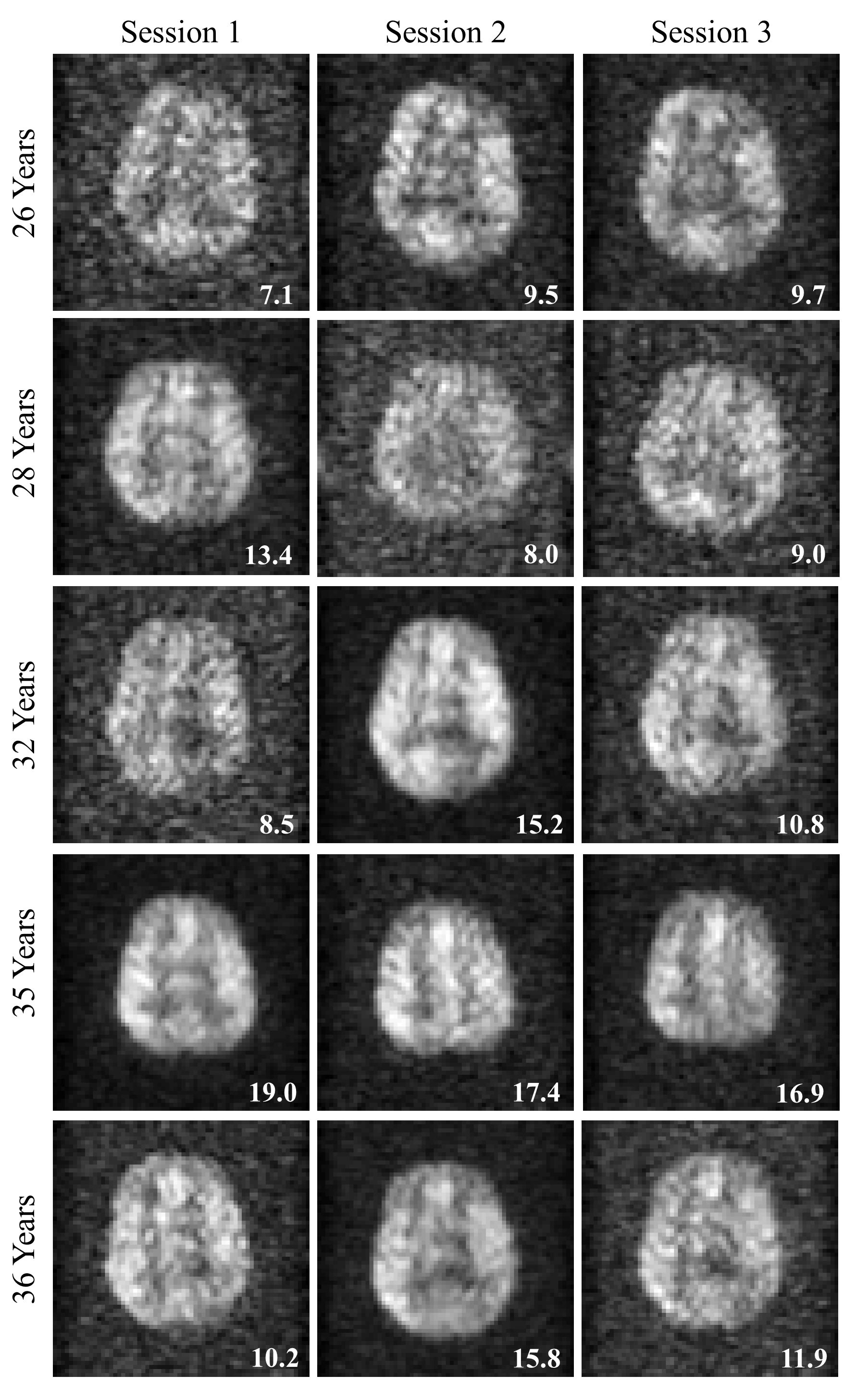
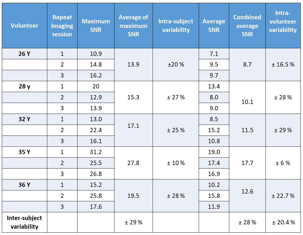
The averaged images from three MR imaging sessions from all the five subjects are shown in Figure 1, and the achievable SNR and variability is provided in Table 1. Over the 5 volunteers, the maximum SNR of 18.8 ± 6.1 and mean SNR of 12.1 ± 3.8 was achievable. The average intra-subject variability of mean SNR was ± 20.4 %.Discussion
The SNR achieved is a function of several factors; (a) polarization of the 129Xe gas, which is at the optimal performance for this application11, (b) sensitivity of the receiver RF coil array, which is optimized in terms of quality factor12 and filling factor2,13, (c) the pulse sequence design and (d) concentration of HP 129Xe in the brain tissue, which is limited here by the 1 L inhaled gas dose. Whilst small gains could be made with further optimization in (a-c) we believe the SNR achieved here at 1.5 T is near optimal level for the 129Xe inhaled dose used (d). Some additional opportunities to improve the SNR would be to investigate ultralow noise amplifiers or imaging at 3.0 T (or higher field) with high density RF coils.
Intra-subject variability (Table 1, ± 6 to 30 %) can be attributed to the environmental variables such as polarization on a particular day, quantity of xenon dose dispensed, T1 relaxation of HP 129Xe gas during the storage after being dispensed and before being administered, concentration of xenon in the lung due to lung inflation state and subject’s physiology (for example, reduced cerebral blood flow due to consumption of caffeine14). Inter-subject variability (Table 1, ± 28 %) can be attributed to variation in resting-state physiology such as lung volume, pulmonary blood volume, pulmonary mean transit time, arterial transit time, cerebral blood volume, cerebral mean transit time and permeability-surface area product of the blood-brain barrier.
Conclusion
This work establishes that the typical mean signal to noise ratio for imaging hyperpolarized 129Xe in the human brain is 12 and typical maximum value is 18 respectively, for a voxel size of 2.8 cm3. The repeatability of the method of ± 20.4 % warrants further investigation as a non-ionizing and injection free means to clinically assess brain tissue perfusion and gas exchange.Acknowledgements
This work was funded by the Engineering and Physical Sciences Research Council (EPSRC - EP/D070252/1), National Institute for Health Research (NIHR - RP-R3-12-027) and Medical Research Council (MRC - MR/M008894/1).References
- Rao M, Stewart NJ, Norquay G, Griffiths PD, Wild JM. High resolution spectroscopy and chemical shift imaging of hyperpolarized 129Xe dissolved in the human brain in vivo at 1.5 tesla. Magn Reson Med 2016;75(6):2227-2234.
- Rao MR, Stewart NJ, Griffiths PD, Norquay G, Wild JM. Imaging Human Brain Perfusion with Inhaled Hyperpolarized 129Xe MR Imaging. Radiology 2018;286(2):659-665.
- Hane FT, Li T, Plata J-A, Hassan A, Granberg K, Albert MS. Inhaled Xenon Washout as a Biomarker of Alzheimer’s Disease. Diagnostics 2018;8(2):41.
- Rao M, Norquay G, Stewart NJ, Hoggard N, Griffiths PD, Wild JM. Assessment of cerebral infarction due to intracranial arterial stenosis in the human brain using hyperpolarized xenon-129 MRI. Proc Intl Soc Mag Reson Med 26 P 3163 2018.
- Swanson SD, Rosen MS, Agranoff BW, Coulter KP, Welsh RC, Chupp TE. Brain MRI with laser-polarized Xe-129. Magnetic Resonance in Medicine 1997;38(5):695-698.
- Swanson SD, Rosen MS, Coulter KP, Welsh RC, Chupp TE. Distribution and dynamics of laser-polarized Xe-129 magnetization in vivo. Magnetic Resonance in Medicine 1999;42(6):1137-1145.
- Zhou X, Sun Y, Mazzanti M, Henninger N, Mansour J, Fisher M, Albert M. MRI of stroke using hyperpolarized 129Xe. NMR Biomed 2011;24(2):170-175.
- Mazzanti ML, Walvick RP, Zhou X, Sun Y, Shah N, Mansour J, Gereige J, Albert MS. Distribution of hyperpolarized xenon in the brain following sensory stimulation: preliminary MRI findings. PloS one 2011;6(7):e21607.
- Latchaw RE, Yonas H, Pentheny SL, Gur D. Adverse reactions to xenon-enhanced CT cerebral blood flow determination. Radiology 1987;163(1):251-254.
- Driehuys B, Martinez-Jimenez S, Cleveland ZI, Metz GM, Beaver DM, Nouls JC, Kaushik SS, Firszt R, Willis C, Kelly KT, Wolber J, Kraft M, McAdams HP. Chronic obstructive pulmonary disease: safety and tolerability of hyperpolarized 129Xe MR imaging in healthy volunteers and patients. Radiology 2012;262(1):279-289.
- Norquay G, Collier GJ, Rao M, Stewart NJ, Wild JM. 129Xe-Rb Spin-Exchange Optical Pumping with High Photon Efficiency. Physical Review Letters 2018;121(15):153201.
- Hayes CE, Edelstein WA, Schenck JF, Mueller OM, Eash M. An efficient, highly homogeneous radiofrequency coil for whole-body NMR imaging at 1.5 T. Journal of Magnetic Resonance (1969) 1985;63(3):622-628.
- Doty FD, Entzminger Jr G, Hauck CD, Staab JP. Practical Aspects of Birdcage Coils. Journal of Magnetic Resonance 1999;138(1):144-154.
- Cameron OG, Modell JG, Hariharan M. Caffeine and human cerebral blood flow: a positron emission tomography study. Life sciences 1990;47(13):1141-1146.
Figures