3316
Visualization of CSF flow of whole brain using 3D dynamic iMSDE SSFP1Department of Radiology, Tokai University Hospital, Isehara, Japan, 2Division of Diagnostic Image Analysis Graduate School of Medicine, Tohoku University, Sendai, Japan, 3Healthcare, Philips Electronics Japan Ltd., Shinagawa, Japan, 4Department of Radiology, Tokai University School of Medicine, Isehara, Japan, 5Course of Electrical and Electronic Engineering, Graduate School of Engineering, Tokai University, Hiratsuka, Japan, 6Department of Neurosurgery, Tokai University School of Medicine, Isehara, Japan
Synopsis
We
reported a technique to visualize the irregular flow of cerebrospinal fluid
(CSF) in whole brain by using dynamic improved motion-sensitized
driven-equilibrium steady-state free precession (dynamic iMSDE SSFP).
The
purpose of this study was to propose a new technique using 3D dynamic iMSDE
SSPF for visualizing the slow and irregular CSF flow of whole brain.
3D dynamic iMSDE SSPF
can visualize the connection between the CSF space and a small lesion. This
technique is suggested to contribute to the diagnosis of various diseases in
the CSF space.
Introduction
We reported a new technique to visualize the irregular flow of cerebrospinal fluid (CSF) by using dynamic improved motion-sensitized driven-equilibrium steady-state free precession (dynamic iMSDE SSFP).1 This sequence has the primary advantages of highlighting various CSF flow in a rapid scan without gating and providing similar visibility to the conventional phase contrast technique. However, because the data in this method can be acquired only in one slice, large region could not be scanned in this method. This method is not useful particularly in case of detecting a connection between the CSF space and a small lesion. To overcome this problem, we here propose a new technique for visualization of CSF flow of whole brain using 3D dynamic iMSDE SSFP. The purpose of this study was to determine basic appropriate parameters and evaluate the feasibility of this technique.Material and Methods
The theory of 3D dynamic iMSDE SSFP is shown in Figure 1. The equipment we used was a 1.5T clinical scanner (Ingenia, Philips, Best, the Netherlands) with either a Torso coil (phantom study) or a dS-Head Spine coil (volunteer study). The basic parameters for dynamic iMSDE SSFP were as follows: 3D balanced TFE with iMSDE, field of view: 250mm, voxel size: 1.2×0.95×2mm, TR/TE: 4.1/2.1 ms, flip angle of 60°, reduction factor: 3 (phase) and 2 (slice), 3D free factor: yes, turbo field echo factor: 300, NSA:1, T2prepTE: 20ms, flow VENC: 1cm/s, direction of MSG: 3 axes, dynamic scan: 8 (MSG-off: 1, MSG-on: 7) and scan time: 2 min 38 s. Phantom study: The flow phantom was made with superabsorbent polymer and tubes with constant flow . We investigated the optimal k-space ordering and acquisition duration time. The signal intensity of flowing water in the tube was measured with varying k-space ordering (linear, low-high) and acquisition duration time (1236, 1649, and 2061 ms) in longitudinal section. Differences of signal intensity for each parameter were assessed by using one-way repeated-measures analysis of variance (ANOVA) with multiple- comparison Dunn's test. Volunteer study: Five healthy volunteers (age range, 25–46 years) were included; written informed consent was obtained from all volunteers. We investigated the optimal acquisition duration time (1236, 2061 ms), as well as CSF flow in the acquired sagittal section and the multi-planar reconstruction (MPR) of the transverse and coronal sections in the volunteers. Visual assessment was performed using a 5-point scale by five radiological technologists. The criteria of the 5-point scale were: 1 (unsatisfactory), 2 (poor image), 3 (fair image), 4 (good image), and 5 (excellent image). Differences of the scores for each parameter were assessed by using the Friedman’s test and multiple-comparison Holm's test.Results
Phantom study k-space ordering: The signal intensity of MSG-on was decreased by low-high ordering, but the signal intensity of MSG-off was also decreased. The more banding artifacts also occurred than linear ordering. Acquisition duration time: signal intensity of MSG-on decreased by shortening the acquisition duration time, but that of MSG-off did not show significant changes (Figure 2, 3). Volunteers study: 3D dynamic iMSDE SSFP with acquisition duration time of 1236 ms achieved significantly higher scores (P <0.05, Figure 4). Transverse and coronal MPR images were able to observe the flow of CSF in the lateral ventricles, the third ventricle, the fourth ventricle, the ventral surface of the brain stem, and cisterna magna (Figure 5).Discussion
Phantom study: Linear ordering and acquisition duration time of 1236 ms were the optimum for this technique. This technique requires signal intensity differences between MSG-off and MSG-on for subtraction. Signal-intensity differences obtained by low-high ordering and shortening of acquisition duration time were larger than those by linear ordering and acquisition duration time of 1236 ms. However, in low-high ordering, the signal intensity of MSG-off was decreased and banding artifacts occurred. Volunteers study: This technique may enable to detect the connection between the CSF space and a small lesion. Detection of slow and irregular flow in CSF required a shortest acquisition duration time. This method features to visualize the CSF flow of the whole brain. It also enables to observe the CSF flow in any cross section.Conclusion
3D dynamic iMSDE SSPF can sensitively detect the slow and irregular CSF flow in whole brain. This technique is suggested to contribute to the diagnosis of various diseases in the CSF space.Acknowledgements
No acknowledgement found.References
1. Horie T, et al. World Neurosurg. 2017; 97(1):523-531.Figures
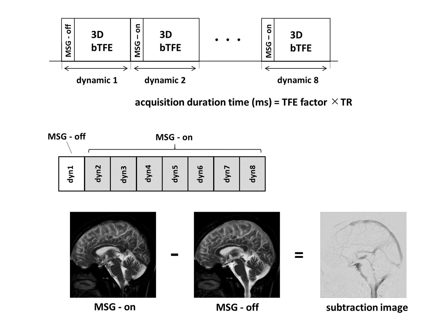
Figure 1 Schematic diagram of the proposed 3D dynamic iMSDE SSFP sequence
3-dimentional balanced turbo field-echo (3DbTFE) acquisitions with and without MSG preparation are dynamically obtained 8 times, whole brain.
To create motion-sensitized contrast, subtraction of the MSG-ON and -OFF images was performed. The first dynamic image without MSG was selected and used for the subtraction.
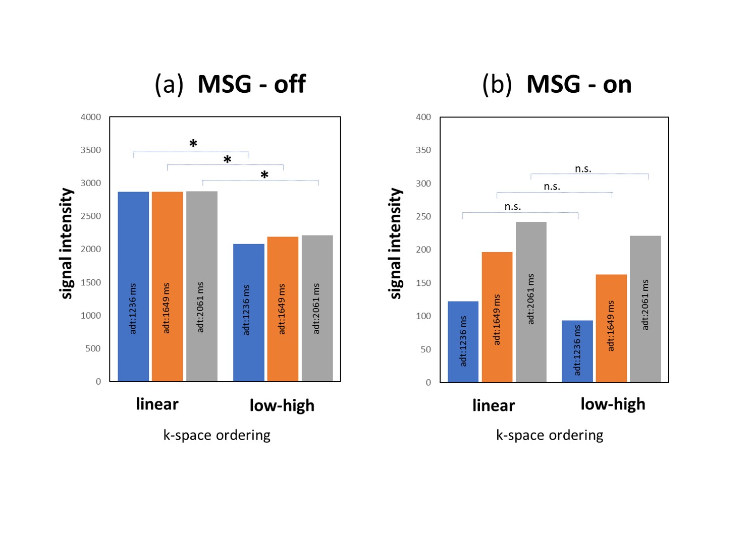
Figure 2 Comparison of the k-space ordering.
(a) Comparison of the k-space ordering in MSG-off. (b) Comparison of the k-space ordering in MSG-on. Values are presented as means ± standard deviation (SD).
* : P<0.05, n.s. : not significant
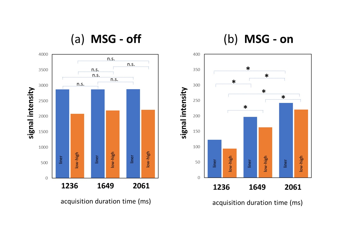
Figure 3 Comparison of the acquisition duration time.
(a) Comparison of the acquisition duration time in MSG-off. (b) Comparison of the acquisition duration time in MSG-on. Values are presented as means ± standard deviation (SD).
* : P<0.05, n.s. : not significant
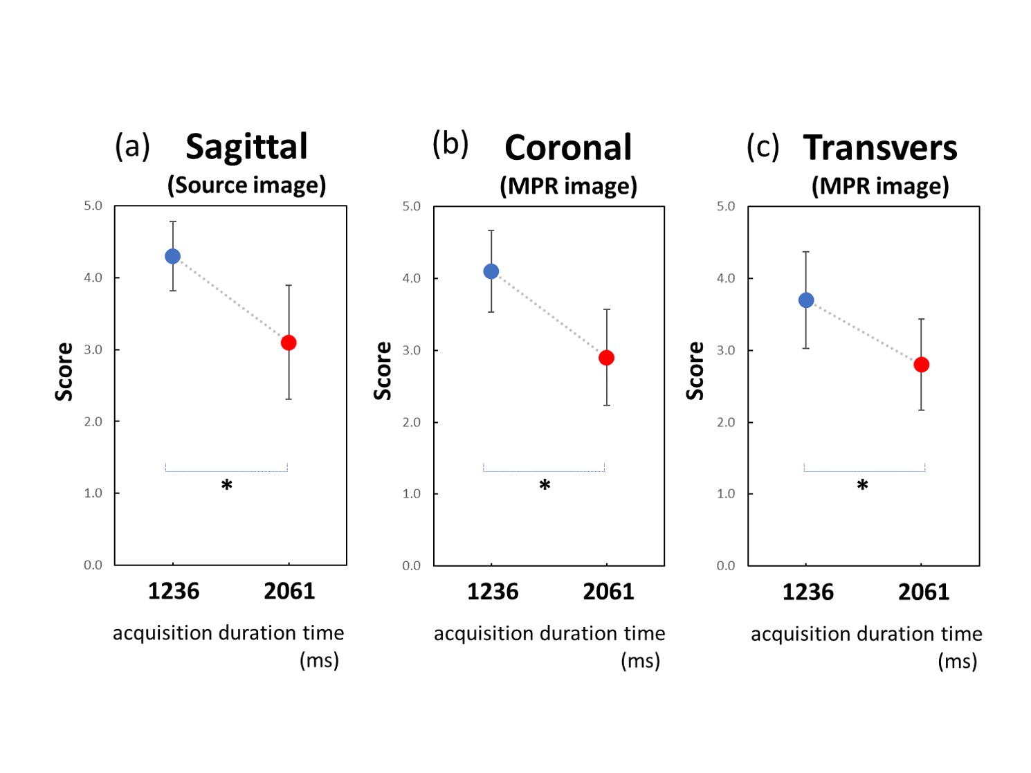
Figure 4 Comparison of the acquisition duration time in volunteers study. (a) Comparison of the acquisition duration time in sagittal image. (b) Comparison of the acquisition duration time in coronal MPR image. (c) Comparison of the acquisition duration time in transvers MPR image. 3D dynamic iMSDE SSFP with acquisition duration time of 1236 ms achieved significantly higher scores (P <0.05).
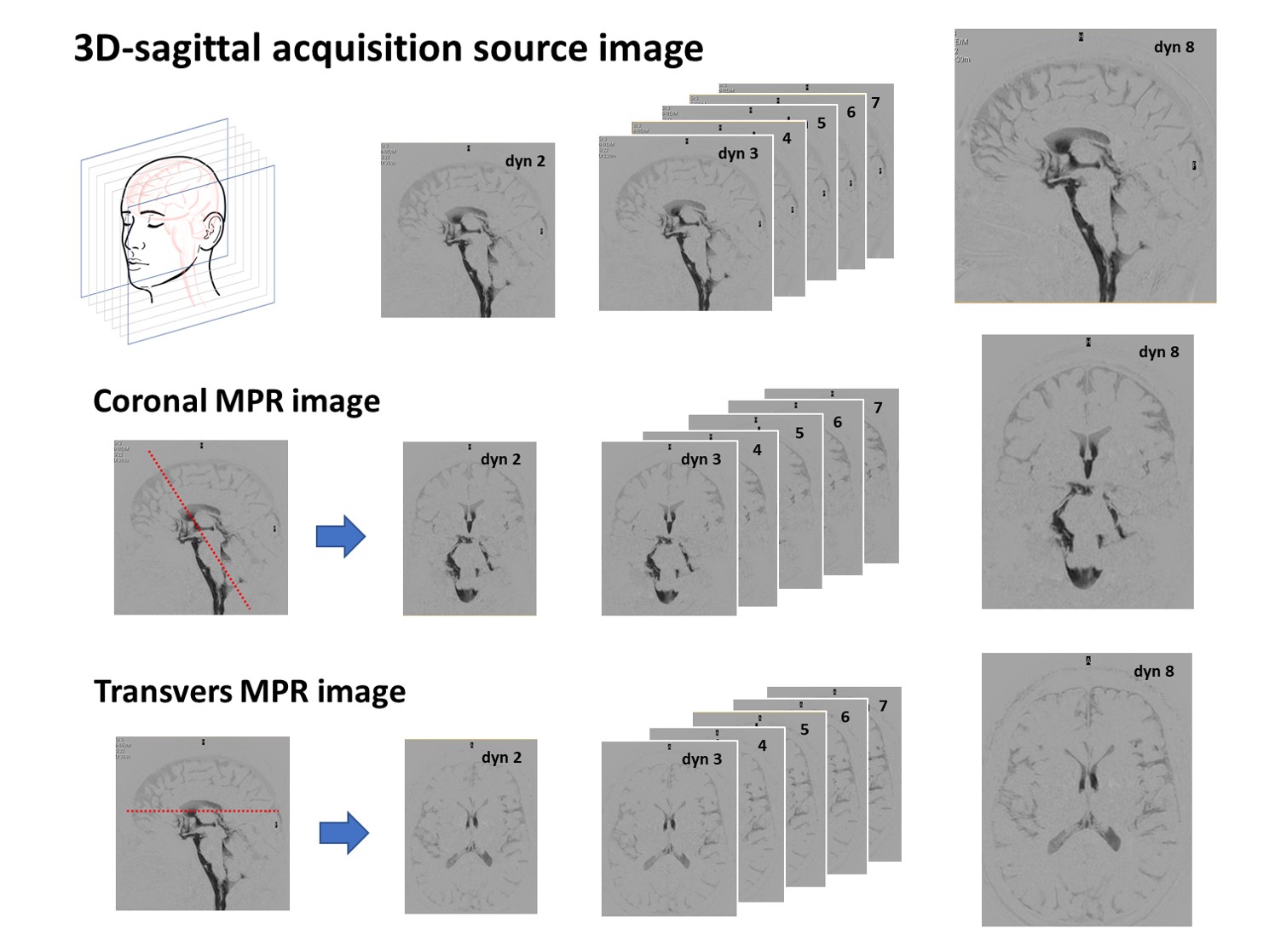
Figure 5 Schematic diagram of Coronal and Transvers MPR image created from 3D dynamic iMSDE SSFP.