3310
Phase opposed cerebral vasoreactivity in multiple sclerosis: evidences of a link between white matter tracts and vascular alterations1I2FH, Institut d’Imagerie Fonctionnelle Humaine, Gui de Chauliac Hospital, Montpellier, France, Montpellier, France, 2Department of Neuroradiology, University Hospital Center, Gui de Chauliac Hospital, France, Montpellier, France, 3LIRMM, Laboratoire d'Informatique, de Robotique et de Microélectronique de Montpellier, France, Montpellier, France, 4Department of Neurology, CHU de Montpellier, Montpellier, France, montpellier, France
Synopsis
Patients with multiple sclerosis (MS) have a higher risk for ischaemic stroke. The current hypothesis states that white matter (WM) fibers alterations causes, through astrocytes, a cerebral vasoreactivity (CVR) disruption resulting in a hypoperfusion. Due to the location of the astrocytes, we expect an altered vasoreactivity mainly around WM tracts. Using a MR vasoreactivity experiment, we could identify altered WM pathways. In MS patients a path from left anterior insula to both precentral gyrus and right middle and superior frontal gyrus highlighted an altered CVR compared to controls. A negative association was found with fNART in the cingulum limbic pathway.
Introduction
Patients with multiple sclerosis (MS) have a higher risk for ischaemic stroke. The current hypothesis states that white matter fibers alterations causes, through astrocytes, a cerebral vasoreactivity (CVR) disruption resulting in a hypoperfusion. Regional differences in vasodilatory capacity can lead to a paradoxical decrease in cerebral blood flow during the hypercapnic challenge. This phenomenon is referred as a blood steal1. It is presumed to be caused by the redistribution of blood from regions in which vasodilation is at a maximum to areas where vasodilation can still occur. In such conditions a decreased BOLD signal can be seen during the challenge. We aimed to test the CVR disruption hypothesis by focusing on blood steal phenomenon during a hypercapnic challenge. Due to the location of the astrocyte, we expect to see such altered vasoreactivity mainly around white matter tracts. To investigate this, we use a simple methodology based on a MR vasoreactivity experiment and hypothesize that the resulting map provides new information compared to other MRI biomarker such as diffusion tensor imaging.
Materials and methods
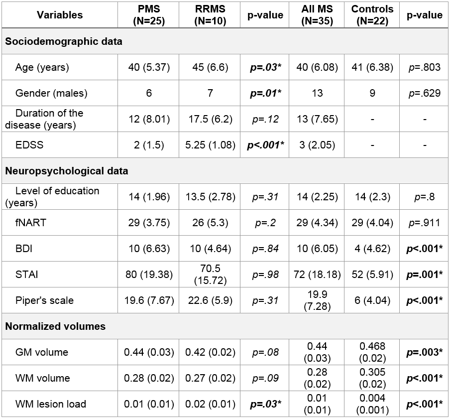
Thirty-five patients with MS (ten progressive and 25 remitting forms) and 22 controls underwent MRI with a hypercapnic challenge to assess cerebral vasoreactivity and a neuropsychological assessment (Fig.1). Blood steal phenomenon is characterized by a decreasing BOLD signal during hypercapnic phase which recovers afterwards. To obtain phase opposed CVR (poCVR), the mean end-tidal carbon dioxide regressor was shifted by one phase in the GLM analysis.
White matter lesions were evaluated from a 3DT1 and a T2FLAIR acquisition. Fractional anisotropy (FA) and mean diffusivity (MD) were estimated using a 30 directions diffusion tensor imaging (DTI). Finally, a time-of-flight angiography was included to rule out any vascular abnormalities. General linear model was used to assess differences between groups and association with clinical parameters. All statistical analysis were adjusted for age, sex and level of education, and a statistical threshold of p<.005 with FWE correction for multiple comparison at cluster level (p<.05) was set for voxel based analysis.Results
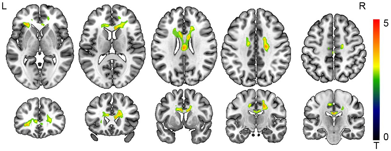
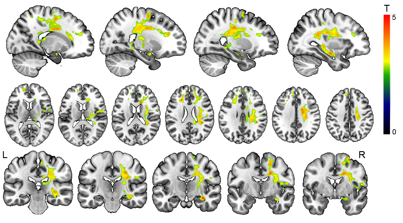
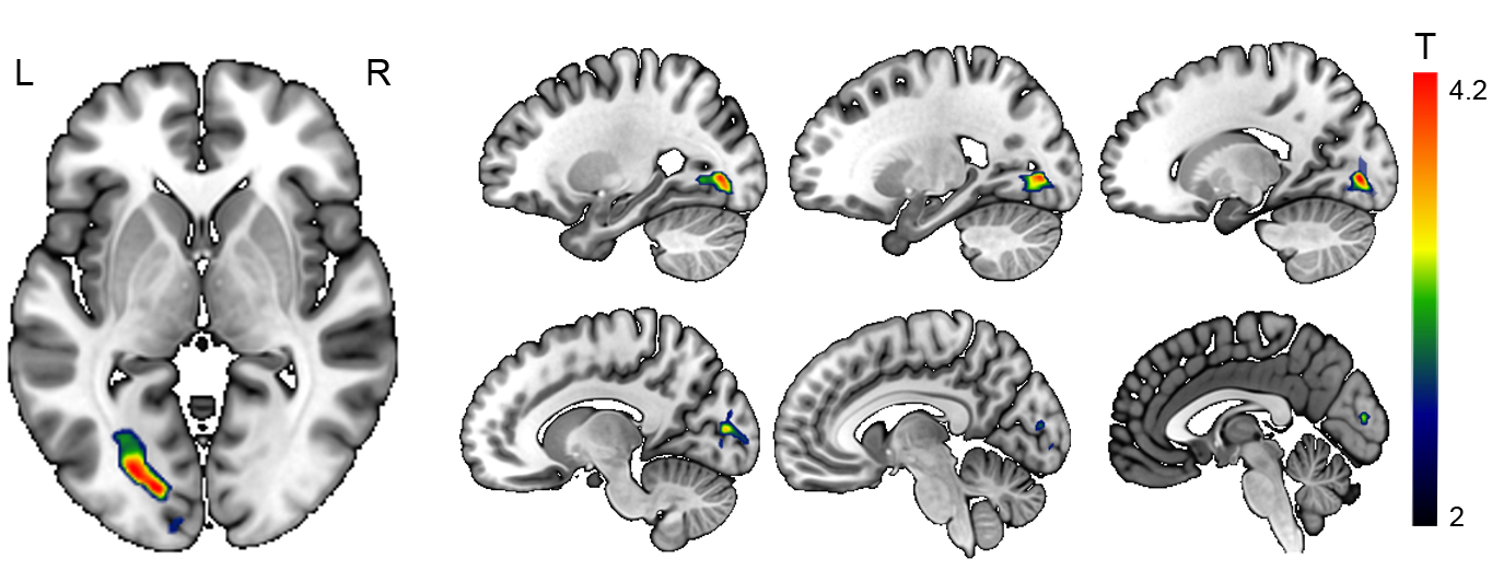
In MS patient, a white matter pathway linking, through anterior part of the corpus callosum, left anterior insula to both precentral gyrus and right middle and superior frontal gyrus was found with higher poCVR compared to controls (Fig.2, P<0.005, cluster P<0.05 FWE corrected). A negative association was found with fNART in a pathway linking right hippocampus to prefrontal cortex through right hemisphere projection fibers (Fig.3, P<0.005, cluster P<0.05 FWE corrected). Progressive MS patients appears to have higher poCVR compared to relapsing-remitting form in left optic radiations and left calcarine cortex (Fig.4, P<0.005, cluster P<0.05 FWE corrected). No association was found with EDSS, cognitive status, depression, fatigue and anxiety score. Average white matter FA were not correlated to poCVR values (Kendall correlation, p=0.99). We didn’t find any FA threshold above or below which values are correlated. The same results were found with MD (Kendall correlation, p=0.68), L1 (Kendall correlation, p=0.97), L2 (Kendall correlation, p=0.72) and L3 (Kendall correlation, p=0.69) maps.Discussion
Alteration of the corpus callosum has been widely described in multiple sclerosis especially in term of fractional anisotropy2, hypoperfusion3,4, and atrophy5. But, rather than a simple focal alteration, we observed an alteration of what appears to be a pathway linking the left insula to the precentral gyrus.
A clear negative association with fNART was found involving prefrontal cortex, projections fibers and right hippocampus. Previous study reported, using FA, that hippocampal–thalamic–prefrontal (cingulum limbic pathway) disruption affects cognitive performance in early RRMS with mild to minimal cognitive impairment6. We were able, based only a vascular characteristic, to observe alterations seemingly following white matter tracts and affecting gray matter (hippocampus). None of the tested DTI parameters was linearly correlated to poCVR values, neither at the voxel level nor at the whole white matter level. It suggests that either those are independant parameters or that DTI measures are less sensitive that poCVR. Of course, we checked only linear correlation between parameters and it remains possible that a nonlinear relationship exists with a correlation appearing only above a certain value of, for example, FA.Conclusion
This study aimed to evaluate the poCVR maps as a potential biomarker for vascular alterations in MS patients. The poCVR maps focuses on voxels with a blood steal profile. We didn’t expect to describe new areas affected in MS, but we hoped to provide new characterization of the well-known alterations. Interestingly, using this vascular feature, most of the results found seem to overlap known fasciculi impacted in MS. This co-occurrence cannot be accidental and suggests a link between axonal loss and vasoreactivity alterations.
Acknowledgements
No acknowledgement found.References
1) Lee, M., Zaharchuk, G., Guzman, R., Achrol, A., Bell-Stephens, T., Steinberg, G.K., 2009. Quantitative hemodynamic studies in moyamoya disease: A review. Neurosurg. Focus 26, E5. https://doi.org/10.3171/2009.1.FOCUS08300
2) Ozturk, A., Smith, S.A., Gordon-Lipkin, E.M., Harrison, D.M., Shiee, N., Pham, D.L., Caffo, B.S., Calabresi, P.A., Reich, D.S., 2010. MRI of the corpus callosum in multiple sclerosis: association with disability. Mult. Scler. Houndmills Basingstoke Engl. 16, 166–177. https://doi.org/10.1177/1352458509353649
3) Filippi, M., Rocca, M.A., 2011. The multiple sclerosis mystery: is there a vascular component? Lancet Neurol. 10, 597–598. https://doi.org/10.1016/S1474-4422(11)70124-7
4) Saindane, A.M., Law, M., Ge, Y., Johnson, G., Babb, J.S., Grossman, R.I., 2007. Correlation of diffusion tensor and dynamic perfusion MR imaging metrics in normal-appearing corpus callosum: support for primary hypoperfusion in multiple sclerosis. AJNR Am. J. Neuroradiol. 28, 767–772.
5) Papathanasiou, A., Messinis, L., Zampakis, P., Papathanasopoulos, P., 2017. Corpus callosum atrophy as a marker of clinically meaningful cognitive decline in secondary progressive multiple sclerosis. Impact on employment status. J. Clin. Neurosci. Off. J. Neurosurg. Soc. Australas. 43, 170–175. https://doi.org/10.1016/j.jocn.2017.05.032
6) Kern, K.C., Gold, S.M., Lee, B., Montag, M., Horsfall, J., O’Connor, M.-F., Sicotte, N.L., 2014. Thalamic–hippocampal–prefrontal disruption in relapsing–remitting multiple sclerosis. NeuroImage Clin. 8, 440–447. https://doi.org/10.1016/j.nicl.2014.12.015
Figures