3294
Feasibility of NODDI for the characterization of multiple sclerosis clinical features1Department of Neurosciences, Biomedicine and Movement sciences, University of Verona, Verona, Italy, 2University Medical Center, Image Sciences Institute, Utrecht, Netherlands, 3Neuroradiology Unit, Department of Diagnostics and Pathology, Verona University Hospital, Verona, Italy, 4Padova Neuroscience Center, University of Padua, Padova, Italy, 5Department of Information Engineering, University of Padua, Padova, Italy, 6Division of Brain Sciences, Faculty of Medicine, Imperial College London Hammersmith Hospital, London, United Kingdom
Synopsis
Neurite Orientation Dispersion and Density Imaging (NODDI) is a diffusion weighted MRI technique introduced to asses neuronal microstructure. NODDI is used to retrieve indexes such as neurite density and fibers orientation dispersion that might be useful to assess neuronal damage and demyelination in multiple sclerosis (MS) MRI images. For this reason, we investigate its ability to differentiate to different MS phenotypes and clinical features in white matter and in multiple areas of the grey matter.
Introduction
Multiple sclerosis (MS) is a chronic inflammatory demyelinating disease causing neurological disability1. There are several phenotypes of MS depending on the symptoms and the pattern of progression of the disease: relapsing-remitting (RR) MS is characterized by the unpredictable alternance of periods of high disease activity and of relative quiescence while primary progressive (PP) MS, instead, presents disability progression from the first symptoms appearance without remission periods. Differences in the disease phenotype and in the clinical outcome of the patients might be related to microstructural changes in brain tissue. Neurite Orientation Dispersion and Density Imaging (NODDI)2 is a novel diffusion weighted (DW) MRI technique introduced to assess the neuronal microstructure morphology changes in a clinically feasible data acquisition setting. Since only a few studies3 investigated the use of this method principally in spinal cord3 and white matter4 (WM) of multiple sclerosis (MS) patients, we explored the differences between NODDI values in the grey matter (GM) and WM of RRMS and PPMS as well as their association with clinical features such as disease duration and Expanded Disability Status Scale (EDSS).Methods
NODDI models the diffusion signal in each voxel as the contribution of three independent compartments: isotropic, intra-neurite and extra neurite compartment as in the following formula:
$$A=(1-F_{iso})(F_{ic}A_{ic}+(1-F_{ic})A_{ec})+F_{iso}A_{iso}$$
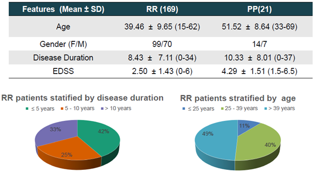
Where $$$A_{ic}$$$, $$$A_{iso}$$$ and $$$A_{ec}$$$ are the intra-cellular, isotropic and extracellular normalized signal fractions and $$$F_{ic}$$$,$$$F_{iso}$$$ and $$$F_{ec}$$$ the corresponding volume fractions2. This model was fitted on data acquired from 169 RR and 21 PP patients of the MS Centre of Verona University Hospital (dataset described in fig.1) to retrieve the images of three tissue indices: Neurite Density Index (NDI), Orientation Dispersion Index (ODI), and isotropic diffusion volume fraction (Fiso), where:
- Fiso accounts for the water free to move such as the interstitial water and Cerebrospinal Fluid (CSF)
- NDI represents the density of neurites estimated from the Fic which represents the water contained in highly anisotropic structures, such as dendrites and axons.
- ODI indicates the level of dispersion of the neurites, low in areas of very coherent fibers, such as in the white matter, and close to 1 where the fibers are very dispersed.
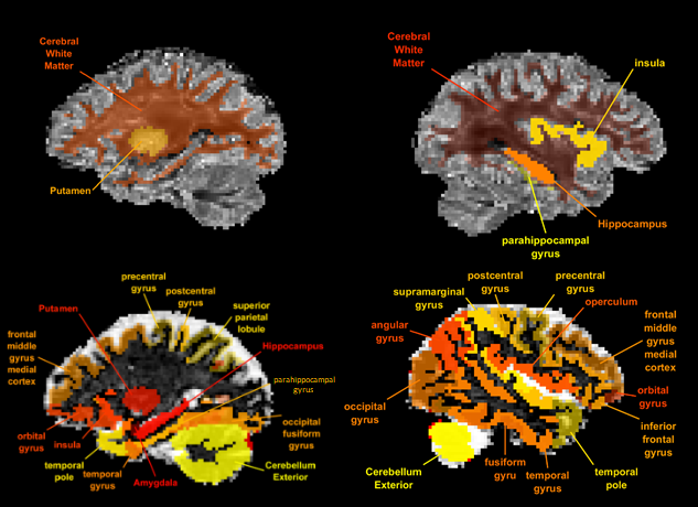
Data acquisition was based on a double shell DW-MRI sequence with 8 non-weighted volumes, 32 gradient directions at b=700s/mm2 and 64 gradients at b=2000s/mm2 for an acquisition time of 20 min. DW-MRI images were preprocessed with Tortoise using a T2w sequence for reference to reduce motion, eddy currents and EPI artifacts. 83 Regions Of Interest (ROIs) were delineated from the T1w data using the Multi Atlas Segmentation with joint Label Fusion (MALF) technique and the Advanced Normalization Tool (ANTs). The T1w data were acquired using a 3D Fast Field Echo with TE/TR 3.7/8.4ms and resolution 1x1x1mm3.
Results
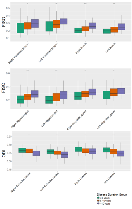
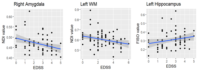
PPMS differed from RRMS in several NODDI parameters: ODI was significantly greater in the PPMS than in the RRMS in both the left and the right WM (p<0.02), in the left and the right Hippocampus (p<0.001, p<0.05, respectively) and in the left and the right Insula (p<0.03, p<0.001, respectively). In addition, ODI was significantly lower in the left Putamen (p<0.01) among PPMS patients, compared to the RRMS (fig.2). $$$F_{iso}$$$ was found to be significantly greater in several cortical regions in the PPMS, compared to the RRMS and in RRMS with long disease duration (>10 years) compared to early RRMS (< 5 years), including in the Cingulate Gyrus, the Hippocampus, the Thalamus and the Insula (p<0.001) (fig.3). Finally, among RR patients, stratified by age (≤25,25-39,>39 years), we observed a negative correlation between EDSS and NDI of the left WM, in the last two age groups only(fig.4).Discussion and Conclusion
Alterations in PPMS
compared to RRMS were observed both in WM and in GM.
Furthermore,
significant difference between RRMS with long disease duration compared to
early RRMS have been detected. Even if further studies are required to
investigate the relation between NODDI and clinical parameters, these data show
that NODDI may represent a sensible tool to assess differential brain microstructure
alterations related to different disease course and evolution.Acknowledgements
No acknowledgement found.References
1. Noseworthy, J. H., Lucchinetti, C., Rodriguez, M. & Weinshenker, B. G. Multiple Sclerosis. N. Engl. J. Med. 2000; 343: 938–952.
2. Zhang, H., Schneider, T., Wheeler-Kingshott, C. A. & Alexander, D. C. NODDI: Practical in vivo neurite orientation dispersion and density imaging of the human brain. Neuroimage 2012; 61: 1000–1016.
3. By, S., Xu, J., Box, B. A., Bagnato, F. R. & Smith, S. A. Application and evaluation of NODDI in the cervical spinal cord of multiple sclerosis patients. NeuroImage Clin. 2017; 15: 333–342.
4. Schneider, T. et al. Sensitivity of multi-shell NODDI to multiple sclerosis white matter changes: a pilot study. Funct. Neurol. 2017; 32: 97–101.
Figures