3293
Axonal Damage in the Optic Radiation Detected by Advanced Diffusion MR Metrics is Associated with Retinal Thinning in Multiple Sclerosis1Department of Radiology, Massachusetts General Hospital / Athinoula A. Martinos Center for Biomedical Imaging, Boston, MA, United States, 2Department of Radiology, Faculty of Medicine, Siriraj Hospital, Mahidol University, Bangkok, Thailand, 3Department of Neurology, Massachusetts General Hospital, Harvard Medical School, Boston, MA, United States
Synopsis
Axonal damage diffusely involves the retina, optic nerve, and optic pathway in MS but lacks a specific imaging biomarker. We investigated the presence of alterations in advanced diffusion MRI metrics derived from WMTI and NODDI in the optic radiation (OR) in MS. We found a significant association between thinning of the retinal nerve fiber layer measured by OCT and reduction in axonal water fraction and intracellular water fraction in the OR as measured by WMTI and NODDI. Our results support the idea that axonal damage is widespread throughout the visual pathway in MS and may be mediated through trans-synaptic degeneration.
Introduction
Damage to the white matter of the visual pathway is common in patients with multiple sclerosis (MS) and may be symptomatic (i.e., optic neuritis) or subclinical. Diffusion tensor imaging (DTI) has been used to demonstrate microstructural damage to the optic radiation (OR) in MS. DTI alterations in the OR relate to retinal thinning in MS, as detected by optical coherence tomography (OCT).1,2 Advanced multi-compartment diffusion MR methods such as white matter tract integrity (WMTI)3,4 and neurite orientation dispersion and density imaging (NODDI)5 may provide greater specificity to axonal integrity. The goal of this study was to determine the presence and extent of changes in WMTI and NODDI metrics in the OR of patients with MS and to investigate the association between these advanced diffusion MRI metrics and retinal thinning as measured by OCT. We hypothesized that WMTI and NODDI metrics would reflect the axonal damage that occurs in the OR in MS, and that axonal alterations revealed by WMTI and NODDI would be associated with retinal thinning.Methods
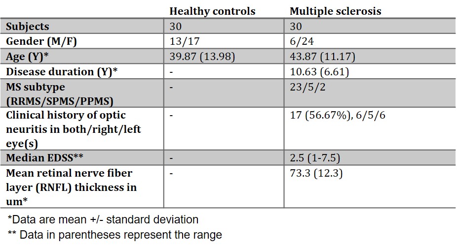
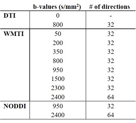
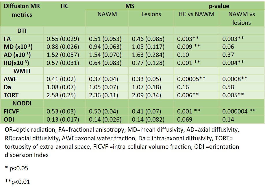
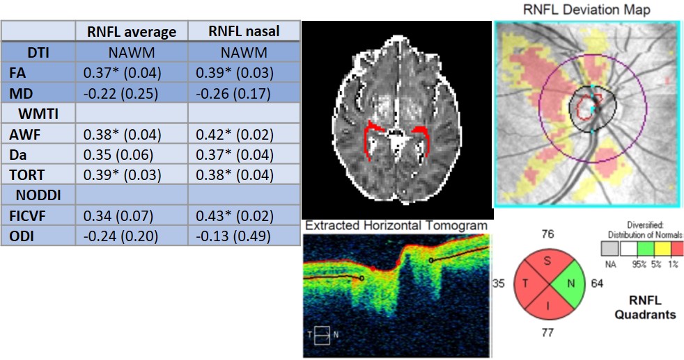
Participants: Thirty MS patients were recruited prospectively from the Massachusetts General Hospital MS Clinic between 2015-2018. Thirty age-matched healthy controls (HC) were also recruited. All MS patients were evaluated with OCT within 30 days of the MRI scan. MRI protocol: All subjects were scanned using a multi-shell diffusion imaging protocol on a dedicated high-gradient 3 Tesla MRI scanner (MAGNETOM CONNECTOM, Siemens Healthineers, Erlangen, Germany) equipped with 300 mT/m maximum gradient strength. The diffusion imaging acquisition parameters were as follows: 2×2×2 mm3 voxels, echo time/repetition time=77/3800 ms, parallel imaging acceleration factor R=2, slice acceleration factor of 2, and anterior-to-posterior phase encoding. Diffusion-encoding parameters for each analysis method are listed in Figure 2. Tract reconstruction: DTI data and probabilistic tractography in FSL were used to reconstruct the courses of both ORs in each participant. FreeSurfer automated cortical segmentation was used to generate regions of interest (ROIs) of the lateral geniculate nucleus (LGN) and V1 of the occipital lobes (Figure 4). The OR region of interest was generated by probabilistic tractography with seed and target regions in the LGN and V1 region. Lesions within the OR were manually segmented and subtracted from the OR to generate normal-appearing white matter (NAWM) masks. Diffusion metrics: Across the voxels that comprised the reconstructed OR, we measured mean values of the following diffusion MRI indices: fractional anisotropy (FA), mean diffusivity (MD), axial diffusivity (AD) and radial diffusivity (RD). WMTI indices of axonal water fraction (AWF), intra-axonal diffusivity (Da), and tortuosity (De,||/De,perp) and NODDI metrics of intracellular volume fraction (FICVF) and orientation dispersion index (ODI) were calculated and reported. Statistical analysis: Differences in diffusion MR metrics between HC and MS patients were assessed by multiple linear regression analysis adjusting for age and sex. Relationships between the OCT data and diffusion MR metrics were assessed using weighted Pearson partial correlation coefficients controlled for age and sex. In this exploratory analysis, we considered p-values<0.05 statistically significant without adjustment for multiple comparisons.Results
Clinical and demographic data for the MS patients and HC are presented in Figure 1. Optic radiation NAWM of MS patients showed significantly decreased FA and increased RD compared to HC, with similar trends seen in MS lesions compared to NAWM (Figure 3). Optic radiation NAWM of MS patients showed significantly decreased AWF and FICVF compared to HC, with similar trends in MS lesions compared to NAWM. Thinning of the RNFL on average and especially in the nasal retinal quadrant correlated with reduction in FA, AWF, Da, tortuosity and FICVF in the NAWM OR. (Figure 4)Discussion and Conclusion
We found a significant reduction in AWF and FICVF within both lesions and NAWM of the OR in MS, as measured by two advanced diffusion MR analysis methods, WMTI and NODDI. Our results suggest that axonal damage may be the underlying substrate of previously observed DTI alterations in the OR. Furthermore, RNFL thickness in the nasal retinal quadrant was specifically correlated with FA, AWF and FICVF in the synaptically connected contralateral OR. The RNFL is composed of unmyelinated axons of the retinal ganglion cells that form the optic nerve and has been proposed as a surrogate marker of axonal damage in MS. Our results support the notion that axonal damage is widespread throughout the visual pathway in MS and may be mediated through trans-synaptic degeneration.Acknowledgements
Funding provided by NIH P41-EB015896, S10-RR019307, K23-NS096056, Conrad N. Hilton Foundation Pilot Innovator Award, and an MGH Claflin Distinguished Scholar Award.References
1. Reich DS, Smith SA, Gordon-Lipkin EM, Ozturk A, Caffo BS, Balcer LJ, et al. Damage to the optic radiation in multiple sclerosis is associated with retinal injury and visual disability. Archives of neurology. 2009;66(8):998-1006
2. Klistorner A, Sriram P, Vootakuru N, Wang C, Barnett MH, Garrick R, et al. Axonal loss of retinal neurons in multiple sclerosis associated with optic radiation lesions. Neurology. 2014;82(24):2165-72
3. Jensen JH, Helpern JA. MRI quantification of non-Gaussian water diffusion by kurtosis analysis. NMR in biomedicine. 2010;23(7):698-710
4. Fieremans E, Jensen JH, Helpern JA. White matter characterization with diffusional kurtosis imaging. NeuroImage. 2011;58(1):177-88
5. Zhang H, Schneider T, Wheeler-Kingshott CA, Alexander DC. NODDI: practical in vivo neurite orientation dispersion and density imaging of the human brain. NeuroImage. 2012;61(4):1000-16
Figures