3292
Neurite orientation dispersion and density imaging predicts disability at 8 years follow-up in relapsing remitting MS patient1Signal Processing Laboratory 5, EPFL, Lausanne, Switzerland, 2AA Martinos Center for Biomedical Imaging, MGH, Harvard Medical School, Charlestown, MA, United States, 3CSEM – Centre Suisse d’Electronique et Microtechnique, Neuchatel, Switzerland, 4Neurologic Clinic and Policlinic, Departments of Medicine, Clinical Research and Biomedical Engineering, University Hospital Basel and University of Basel, Basel, Switzerland, 5Translational Imaging in Neurology (ThINk) Basel, Department of Medicine and Biomedical Engineering, University Hospital Basel and University of Basel, Basel, Switzerland, 6Departement of Neurology, Centre Hospitalier Universitaire Vaudois (CHUV) and University of Lausanne (UNIL), Lausanne, Switzerland
Synopsis
Neurite orientation dispersion and density imaging (NODDI) is a diffusion imaging technique that uses diffusion gradients of different strengths to provide novel metrics of axonal and dendrites integrity. In this study, we explored the value of NODDI metrics - in lesions and normal appearing tissue - to predict the long term disability in relapsing-remitting MS (RRMS) patients. NODDI metrics in NAWM and lesions showed significant correlations with patients disability at 8 years follow-up. Future studies should explore the predictive value of NODDI metrics in MS lesions and in larger cohorts of MS patients.
Introduction
Multiple sclerosis (MS) is a chronic inflammatory and
degenerative disease of the central nervous system characterized by the
presence of focal lesions and diffuse demyelination, gliosis and axonal damage
in non lesional “normal-appearing” tissue1. Neurite
orientation dispersion and density imaging (NODDI)
is a recent diffusion imaging technique that uses diffusion gradients of
different strengths to provide novel metrics of axonal and dendrites integrity2.
Previous cross-sectional works showed that NODDI is sensitive to detect brain
structural damage in MS patients compared to controls3-5. In this
study, we explore the value of NODDI metrics - in lesions and normal appearing
tissue - to predict the long-term disability in relapsing-remitting MS (RRMS) patients.Methods
Thirty early RRMS patients with less than 5 years
from initial symptoms
[10 males, 34.6(8.4) mean age(standard deviation)] were recruited at the University Hospital of
Lausanne (CHUV) and scanned on a 3T Siemens Prisma scanner (32-head channel
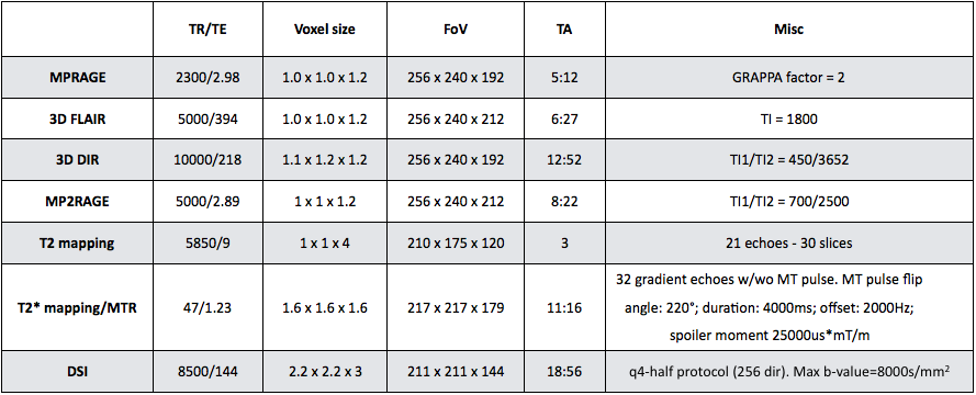
coil) (see Figure 1 for scanning sequences and parameters). Patients had mean Expanded
Disability Status Scale (EDSS) of 1.5[1-2] (median[range]) at baseline and EDSS
of 1.5[1.5-3] (median[range]) at 8 years follow-up.
Mood and fatigue were evaluated using the Hospital
Anxiety and Depression scale (HAD, HADA and HADD for anxiety and depression, resp.). Diffusion weighted images (DWI) were denoised using6, corrected for eddy currents7 and fitted to the NODDI model8. T1 maps were derived from the
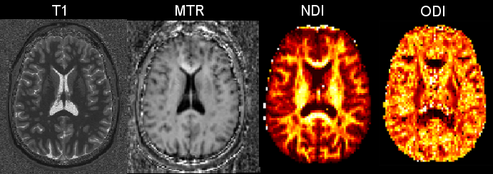
MP2RAGE volume as in9. MTR maps were derived from M0
and MT T2* data9 (see Figure 2). All maps were registered to the
non-weighted diffusion image (b0) using Elastix10. Regions of interest (ROI) were
automatically extracted from the MPRAGE image using an in-house software based
on variational expectation-maximization tissue classification11. Lesions were manually counted
by two expert raters by consensus on 3D FLAIR, 3D DIR and MP2RAGE images and a
lesion mask was obtained by merging the masks on each single contrast as
previously performed12-14.
The non-lesion normal appearing white matter (NAWM) was then segmented in
parietal, frontal occipital and temporal regions of interest (ROIs). In each NAWM ROI and in lesions, mean NODDI parameters (neurite density
index-NDI and orientation dispersion index – ODI) as well as mean T1 and MTR vlaues were computed. A generalized model (GLM)
with step-wise regression was used to predict the disability score (EDSS)
8-years after baseline using MRI metrics, age and gender, as well as HADA and HADD as covariates. All
data was adapted using the Box-Cox power transformation to satisfy the models
assumption for normality.Results
In lesions, GLM analysis showed significant correlation between EDSS at 8-years follow-up and (i) NDI mean values in lesions and (ii) depression scores (HADD) (adj-R2=0.56, corrected p-value=0.04). In normal appearing WM, GLM analysis showed significant correlation between EDSS at 8-years follow-up and (i) NDI mean values in the parietal WM and (ii) ODI mean values in the temporal WM as well as (iii) gender (adj-R2=0.4, corrected p-value = 0.02).Discussion and Conclusion
We applied a generalized linear model on NODDI,
T1 and MTR metrics in NAWM at baseline in our cohort of RRMS patients to
predict EDSS scores at 8 yeas follow-up. NODDI
metrics in NAWM – and not T1 and MTR metrics – showed significant correlations
with patients’ disability. NODDI is a recent diffusion imaging technique that
uses diffusion gradients of different strengths to model and quantify axonal
and dendrites integrity2, which allows the characterization of the neurite
integrity in non-lesional tissue. Future studies should explore the predictive
value of NODDI metrics in MS lesions and in larger cohorts of MS patients.Acknowledgements
This study was supported by the Swiss National Science Foundation, under grants P2ELP2_172286 to EF, P2LAP3_164894 to GB and PP00P3_176984 to CG. The funding source had no role in study design; in the collection, analysis, and interpretation of data; in the writing of the report or in the decision to submit for publication.References
[1] Noseworthy JH, Lucchinetti C, Rodriguez M, Weinshenker BG. Multiple sclerosis. N Engl J Med. Sep 28; 343(13):938-52, 2000.
[2] Zhang H, Schneider T, Wheeler-Kingshott CA, Alexander DC (2012). NODDI: practical in vivo neurite orientation dispersion and density imaging of the human brain. NeuroImage, 2017; 61(4), 1000–16.
[3] Granberg T, Fan Q, Treaba CA, Ouellette R, Herranz E, Mangeat G, Louapre C, Cohen-Adad J, Klawiter EC, Sloane JA, Mainero C. In vivo characterization of cortical and white matter neuroaxonal pathology in early multiple sclerosis. Brain. 2017 Nov 1;140(11):2912-2926. doi: 10.1093/brain/awx247.
[4] By S, Xu J, Box BA, Bagnato FR, Smith SA. Application and evaluation of NODDI in the cervical spinal cord of multiple sclerosis patients. Neuroimage Clin. 2017 May 17;15:333-342.
[5] Grussu F, Schneider T, Tur C, Yates RL, Tachrount M, Ianuş A, Yiannakas MC, Newcombe J, Zhang H, Alexander DC, DeLuca GC, Gandini Wheeler-Kingshott CAM. Neurite dispersion: a new marker of multiple sclerosis spinal cord pathology? Ann Clin Transl Neurol. 2017 Aug 15;4(9):663-679.
[6] St-Jean S et al. Non Local Spatial and Angular Matching: Enabling higher spatial resolution diffusion MRI datasets through adaptive denoising. Medical Image Analysis, 2016; 32:115 – 130
[7] https://fsl.fmrib.ox.ac.uk/fsl
[8] Daducci A, Canales-Rodríguez EJ, Zhang H, Dyrby TB, Alexander DC, Thiran J-P. Accelerated Microstructure Imaging via Convex Optimization (AMICO) from diffusion MRI data. Nueorimage, 2015; 105:32-44 [
9] Bonnier G, Roche A, Romascano D, Simioni S, Meskaldji DE, Rotzinger D, Lin Y-C, Menegaz G, Schluep M, Du Pasquier R, Sumpf TJ, Frahm J, Thiran J-P, Krueger G, Granziera C. Advanced MRI unravels the nature of tissue alterations in early multiple sclerosis. Annals of Clinical and Translational Neurology, 2014; 1(6):423-432
[10] S. Klein, M. Staring, K. Murphy, M.A. Viergever, J.P.W. Pluim. elastix: a toolbox for intensity based medical image registration, 2010 IEEE Transactions on Medical Imaging, 29(1):196 - 205
[11] Roche A, Ribes D, Bach-Cuadra M, Kruger G. On the convergence of EM-like algorithms for image segmentation using Markov random fields. Med Image Anal 2011;15:830–839
[12] Bonnier G, Roche A, Romascano D, Simioni S, Meskaldji DE, Rotzinger D, Lin Y-C, Menegaz G, Schluep M, Du Pasquier R, Sumpf TJ, Frahm J, Thiran J-P, Krueger G, Granziera C. Multicontrast MRI Quantification of Focal Inflammation and Degeneration in Multiple Sclerosis BioMed Research International, 2015.
[13] Bonnier G, Kober T, Schluep M, Du Pasquier R, Krueger G, Meuli R, Granziera C, Roche, A. A New Approach for Deep Gray Matter Analysis Using Partial-Volume Estimation, Plos One, 2016, 11(2): e0148631
[14] Bonnier G, Maréchal B, Fartaria MJ, Falkowskiy P, Marques JP, Simioni S, Schluep, M, Du Pasquier R, Thiran, J-P, Krueger G, Granziera C. The Combined Quantification and Interpretation of Multiple Quantitative Magnetic Resonance Imaging Metrics Enlightens Longitudinal Changes Compatible with Brain Repair in Relapsing-Remitting Multiple Sclerosis Patients. Frontiers in neurology, 2017, 8:506
Figures