3290
Sensitivity of NODDI and two-compartment SMT parameter maps in multiple sclerosis1Queen Square MS Centre, Department of Neuroinflammation, UCL Queen Square Institute of Neurology, Faculty of Brain Sciences, University College London, London, United Kingdom, 2Croydon University Hospital, London, United Kingdom, 3Centre for Medical Image Computing, Department of Computer Science, University College London, London, United Kingdom, 4Department of Medical Physics and Biomedical Engineering, University College London, London, United Kingdom, 5Centre for Medical Image Computing, Medical Physics and Biomedical Engineering, University College London, London, United Kingdom, 6Universitat Oberta de Catalunya, Barcelona, Spain, 7NIHR UCLH Biomedical Research Centre, London, United Kingdom, 8Department of Brain and Behavioural Sciences, University of Pavia, Pavia, Italy, 9Brain MRI 3T Research Centre, IRCCS Mondino Foundation, Pavia, Italy
Synopsis
We make a novel comparison of two diffusion MRI techniques
modelling white matter microstructure: neurite orientation dispersion and
density imaging (NODDI) and spherical mean technique (SMT) in 63 Multiple
Sclerosis (MS) patients and 28 healthy controls using tract based spatial
statistics. Both techniques show that there is a reduction in the intracellular
volume fraction and an increase in neurite orientation dispersion in lesions
and normal appearing white matter in MS patients when compared to controls. Additionally,
SMT appears more sensitive to these differences, identifying a larger number of
voxels showing significant differences between patients and controls in the
studied parameters.
Introduction
Diffusion tensor imaging (DTI) is an MRI technique used to detect and characterise tissue damage beyond focal lesions in diseases including multiple sclerosis (MS)1. However, DTI metrics are surrogate indices lacking direct biological interpretation, and the assumption of Gaussian diffusion is often invalid in highly organised, multi-compartment white matter tissue2. Recent techniques such as neurite orientation dispersion and density imaging (NODDI)3 and the spherical mean technique (SMT)4,5 try to overcome DTI’s limitations, while providing metrics with a direct biophysical interpretation, using clinically feasible scanning protocols.
NODDI and SMT provide metrics with similar biological interpretations. However, it is currently unclear which offers the higher sensitivity to tissue damage in MS. In this work we directly compare these two techniques, and study with tract based spatial statistics (TBSS) their ability to discriminate between differences in intracellular volume fraction and orientation dispersion between MS patients and healthy controls, in lesions and normal appearing white matter (NAWM).
Methods
63 patients with relapsing remitting MS (15 men, age: 47±8y) were compared with 28 healthy controls (9 men, age: 35±10y). All subjects were scanned with a 3T Philips Achieva MRI system. Scanning protocol: multi-shell diffusion scans (resolution: 2.5x2.5x2.5mm; TE=82ms; TR=14s); anatomical PD-T2 images for MS lesion outlining (resolution: 1x1x3mm, TE=10ms, TR=625ms).
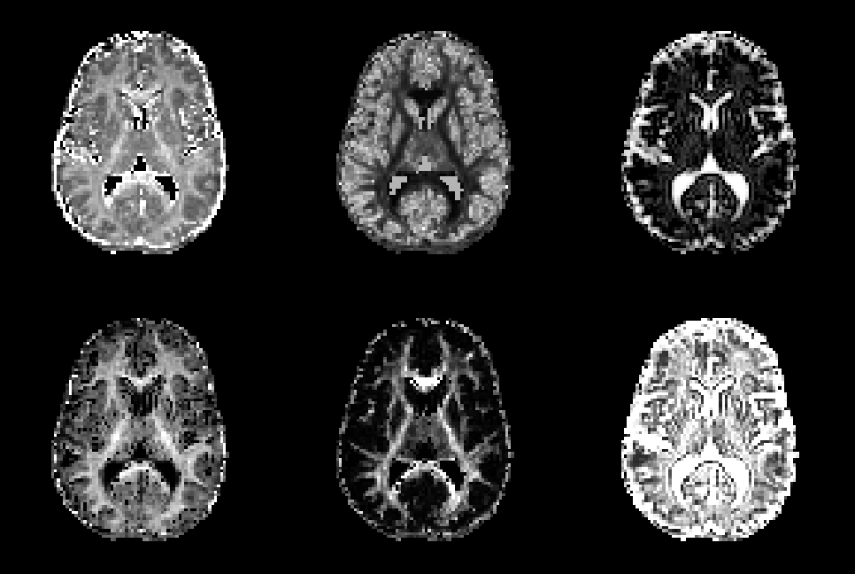
Diffusion scans were corrected for motion and eddy current distortions with FSL’s eddy tool6. Subsequently, NODDI and SMT metrics were calculated using the NODDI toolbox7 for MATLAB8 and the freely available SMT code9. This provides the following metrics: for NODDI, orientation dispersion index (ODI), isotropic volume fraction and intracellular volume fraction (fic-NODDI); for SMT, orientation dispersion entropy (ODE), intrinsic neural diffusivity and intracellular volume fraction (fic-SMT). Sample images are shown in Figure 1.
Comparison was made with TBSS using standard tools in the FMRIB diffusion toolbox (FDT) in FSL10,11,12. Diffusion scans were processed with DTI to obtain fractional anisotropy (FA) images, which were aligned to a standard space and a mean image taken. This image was ‘skeletonised’ by suppressing non-maximal FA voxels perpendicular to local tract structure. Each subject’s aligned FA image was projected onto this skeleton. The mappings used to create the skeletonised FA image were used to create standardised, skeletonised images of the metrics listed above (ODI, fic-NODDI, fiso, ODE, fic-SMT, diffusivity).
Lesion masks were used to classify the skeleton into NAWM, where no lesions were present, and areas where the lesion density was >5% and hence the influence of lesions on the metrics could be considered significant. Lesions were segmented manually on the PD-T2 images and co-registered non-linearly to the TBSS space using warping transformations provided by the TBSS pipeline.
Control and patient images were compared voxelwise with a two-sample t-test. This was performed using FSL randomise13. 10,000 permutations were used. A two-tailed significance level of 5% was used using threshold-free cluster enhancement, correcting for multiple comparisons.
Results
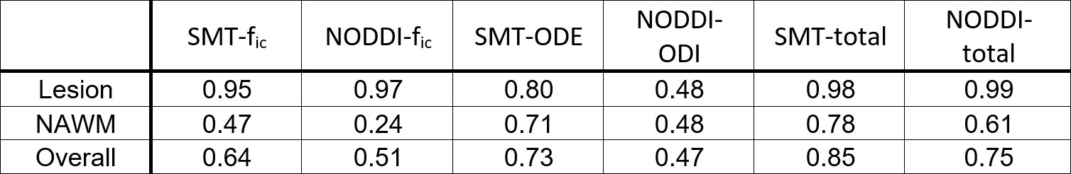
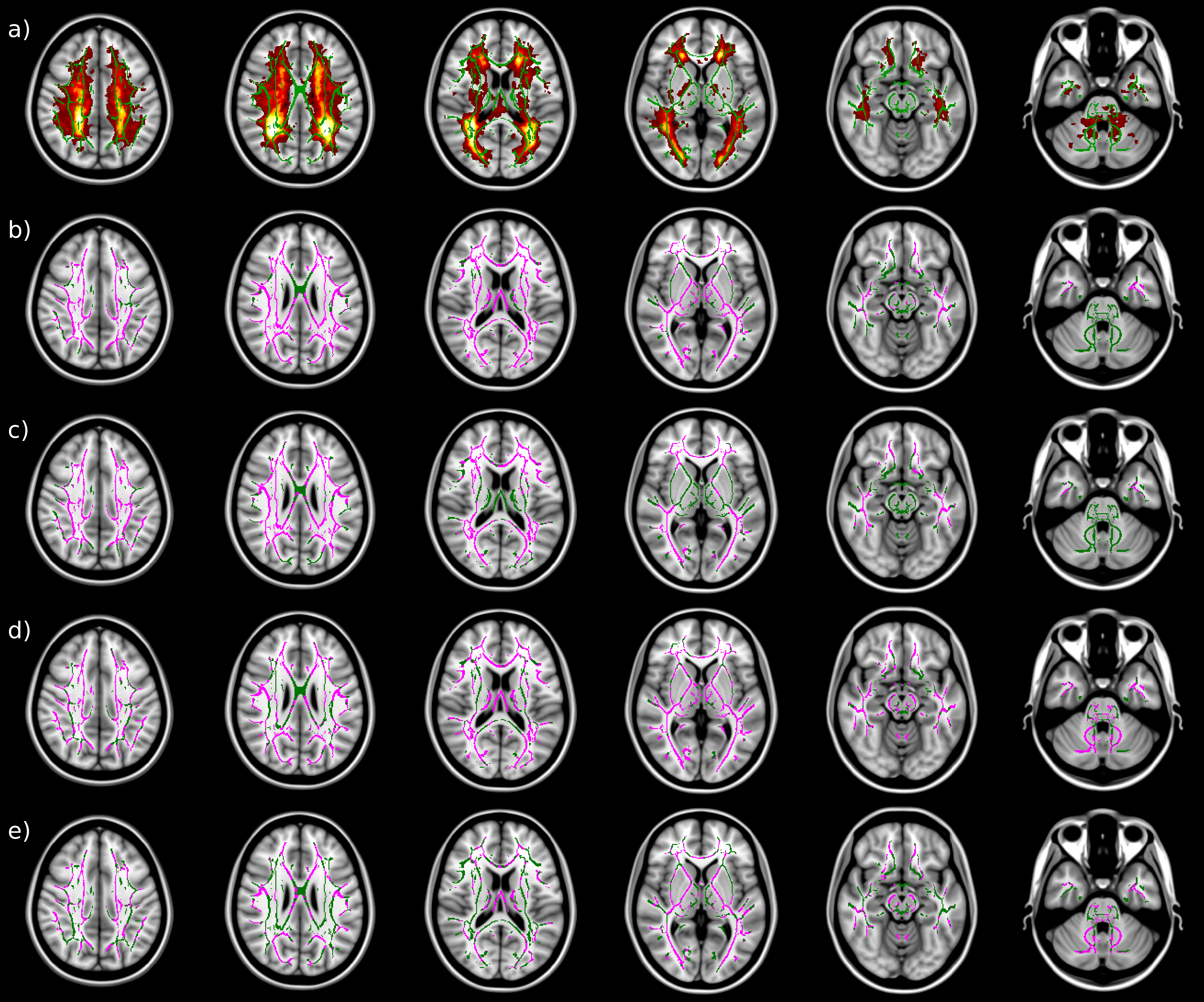
MS patients show a lower intracellular volume fraction than controls over most of the WM skeleton (figures 2 and 3). This was particularly true in lesions with both metrics finding significant differences in >95% of lesional voxels. The extent of the discriminative voxels was greater for fic-SMT than for fic-NODDI.
MS patients showed higher orientation dispersion than controls in both NAWM and lesioned areas using both SMT and NODDI. The extent of discriminative voxels was larger for SMT than NODDI.
Discussion
We find an increase in orientation dispersion in lesions and NAWM using both NODDI and SMT. Previous studies on MS-related changes in ODI in lesions and NAWM are not in agreement14,15. On the other hand, the finding of lower intracellular volume fraction in both lesions and NAWM in MS patients than controls agrees with current knowledge14-16. Here, we extend previous results by showing a consistency between NODDI and SMT. Given TBSS’s lower reliability around lesions, resulting from the decreased FA seen within them, the conclusion for NAWM appears the more robust, and provides evidence that the orientation dispersion of neurites does increase in NAWM in MS.
The results from SMT and NODDI are, in broad terms, highly comparable. Although both observe increases in neurite orientation dispersion and reduced intracellular volume in both lesional and NAWM voxels, our data favours SMT as a more sensitive test for discriminating between MS patients and controls.
Conclusion
Both NODDI and SMT are sensitive in detecting differences in neurite density and orientation dispersion between MS patients and controls. While in areas with a significant lesional burden the two techniques provide similar results, in NAWM, when differentiating MS patients from controls, there is a clear advantage in the sensitivity of SMT over NODDI.Acknowledgements
This project has received funding from the European Union’s Horizon 2020 research and innovation programme under grant agreement No. 634541 and No. 666992.. Engineering and Physical Sciences Research Council (EPSRC EP/R006032/1, M020533/1, G007748, I027084, M020533, N018702). Spinal Research (UK), Wings for Life (Austria), Craig H. Neilsen Foundation (USA) for INSPIRED. UK Multiple Sclerosis Society (grants 892/08 and 77 year 2017). Department of Health's National Institute for Health Research Biomedical Research Centres (BRC R&D 03/10/RAG0449). Guarantors of Brain post‐doctoral non‐clinical fellowships.References
[1] Sbardella E, Tona F, Petsas N, Pantano P. (2013) DTI Measurements in Multiple Sclerosis: Evaluation of Brain Damage and Clinical Implications. Mult. Scler. Int. 2013;2013:671730.
[2] Alexander D, Barker G and Arridge S. (2002). Detection and modeling of non‐Gaussian apparent diffusion coefficient profiles in human brain data. Magn. Reson. Med., 48: 331-340.
[3] Zhang H, Schneider T, Wheeler-Kingshott CA, Alexander DC. (2012). NODDI: practical in vivo neurite orientation dispersion and density imaging of the human brain. Neuroimage. 61(4):1000-16.
[4] Kaden E, Kruggel F, Alexander DC. (2015). Quantitative mapping of the per-axon diffusion coefficients in brain white matter. Magn Reson Med. 75(4):1752-63.
[5] Kaden E, Kelm ND, Carson RP, Does MD, Alexander DC. (2015). Multi-compartment microscopic diffusion imaging. Neuroimage. 139:346-359.
[6] Andersson J and Sotiropoulos S. (2016). An integrated approach to correction for off-resonance effects and subject movement in diffusion MR imaging. NeuroImage, 125:1063-1078.
[7] NODDI Matlab toolbox [updated 11/08/2017, cited 04/11/2018]. Availible from: http://mig.cs.ucl.ac.uk/index.php?n=Tutorial.NODDImatlab)
[8] The MathWorks, Inc., Natick, Massachusetts, United States
[9] SMT [cited 04/11/2018]. Available from: https://github.com/ekaden/smt
[10] Behrens T, Woolrich M, Jenkinson M, Johansen-Berg H, Nunes R, Clare S, Matthews P, Brady J, and Smith S. (2013) Characterization and propagation of uncertainty in diffusion-weighted MR imaging. Magn. Reson. Med. 50(5): 1077-1088.
[11] Behrens T, Johansen-Berg H, Jbabdi S, Rushworth M, and Woolrich M. (2007) Probabilistic diffusion tractography with multiple fibre orientations. What can we gain? NeuroImage, 23:144-155.
[12] Smith S, Jenkinson M, Johansen-Berg H, Rueckert D, Nichols T, Mackay C, Watkins K, Ciccarelli O, Cader M, Matthews P, and Behrens T. (2006) Tract-based spatial statistics: Voxelwise analysis of multi-subject diffusion data. NeuroImage, 31:1487-1505.
[13] Winkler A, Ridgway G, Webster M, Smith S, Nichols T. (2014) Permutation inference for the general linear model. NeuroImage. 92:381-397.
[14] Schneider T, Brownlee W, Zhang H, Ciccarelli O, Miller DH, Wheeler-Kingshott CG. (2017) Sensitivity of multi-shell NODDI to multiple sclerosis white matter changes: a pilot study. Funct. Neurol. 32(2):97-101.
[15] De Santis S, Bastiani M, Droby A, Kolber P, Zipp F, Pracht E, Stoecker T, Groppa S, Roebroeck A. (2018) Characterizing Microstructural Tissue Properties in Multiple Sclerosis with Diffusion MRI at 7 T and 3 T: The Impact of the Experimental Design. Neuroscience. 2018 Apr 7.
[16] Granberg T, Fan Q, Treaba C, Ouellette R, Herranz E, Mangeat G, Louapre C, Cohen-Adad J, Klawiter E, Sloane J, Mainero C. (2017). In vivo characterization of cortical and white matter neuroaxonal pathology in early multiple sclerosis. Brain. 140(11):2912-2926.
Figures