3289
Altered whole-brain functional networks associated with cognitive dysfunction in end-stage renal disease patients with maintenance hemodialysis1Imgaing department, NO.215 Hospital of Shaanxi Nuclear Industry, Xianyang, China, 2Medical Imaging department, The First Affilicated Hospital of Xi'an Jiaotong University, Xi'an, China, 3Imgaing department, The First Affilicated Hospital of Xi'an Jiaotong University, Xi'an, China
Synopsis
We focused on exploring the whole-brain resting-state functional networks abnormality by using the rs-fMRI with independent component analysis (ICA) algorithm and the relationships with cognitive dysfunction in ESRD patients with hemodialysis. 24 maintenance hemodialysis patients (MHD group) and 26 healthy control subjects (HCs) were evaluated a battery of neuropsychological tests and rs-fMRI scans. Compared with HCs, the MHD group showed worse neuropsychological performances and related decreased functional connectivity in the default mode network, the right frontoparietal network, and the sensorimotor network. Our study might contribute to better understanding underlying neuropathological substrate of cognitive dysfunction in patients with ESRD.
Introduction
As the most serious stage of chronic kidney disease (CKD), end-stage renal disease (ESRD) 1 is characterized by multi-organ dysfunction and defined as when renal failure deteriorate to an estimated glomerular filtration rate (eGFR) less than 15 ml/min/1.73 m2. In the United States, approximately 8% of the population is disturbed by CKD, and 571,000 ESRD patients receive maintenance hemodialysis or peritoneal dialysis treatment2. Cognitive dysfunction3, as well as sensorimotor abnormality4 and affective disturbance5, are considered to have a significant influence on the therapeutic regimen and dietary restriction in ESRD patients and associated with hospitalization, disability, and death6. Recently, more attention has been focused on investigating cognitive dysfunction and related changed intrinsic brain function in ESRD patients by using resting-state functional magnetic resonance imaging (rs-fMRI) 6-8. Nevertheless, to our knowledge, few researches investigated the underlying neuropathological substrate at the whole-brain network perspective in ESRD patients. Here, We focused on exploring the whole-brain resting-state functional networks abnormality by using the rs-fMRI with independent component analysis (ICA) algorithm and the relationships with cognitive dysfunction in ESRD patients with hemodialysis.Methods
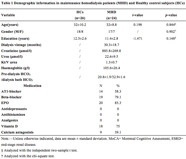
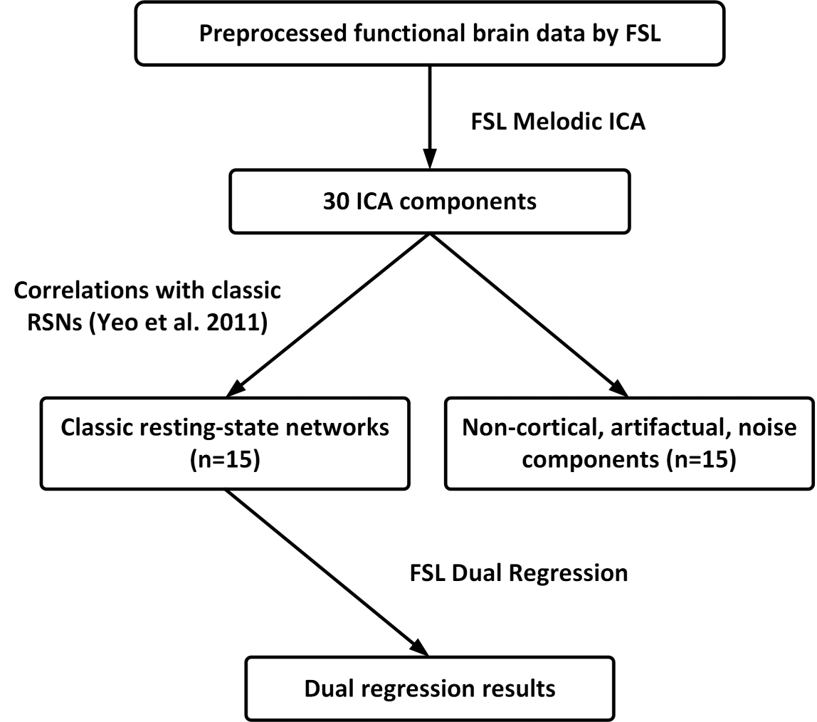
24 maintenance hemodialysis patients (MHD group) (mean age 32±8.8 years) and 26 healthy control subjects (HCs) (mean age 32±10.2 years) were included in our study (Figure 1). A GE 3.0T Discovery 750 scanner with an 8-channel phase array head coil obtained the MRI data. Individual high-resolution T1-weighted anatomical images were collected using a 3D Brain Volume imaging sequence (BRAVO). All the participants were evaluated a battery of neuropsychological tests including the Montreal Cognitive Assessment (MoCA), Auditory Verbal Learning Test (AVLT), Trail Making Test A (TMT-A), and a computed based n-back working memory battery. We also assessed the affective Status and Sleep Condition by using Beck Depression Inventory (BDI), Beck Anxiety Inventory (BAI), and Pittsburgh Sleep Quality Index (PSQI). We collected the laboratory blood tests and clinical information in all ESRD patients, including the serum creatinine, urea, hemoglobin; pre-dialysis HCO3/dialysis bath HCO3, and minimum kinetic transfer/volume urea measurement (Kt/V urea). No laboratory blood tests were performed for the healthy control subjects. FSL build 5.06 was performed on all processing of brain data9. Independent Components Analysis (ICA)10 was performed on the preprocessed functional brain data using Melodic ICA version 3.14 (Figure 1).Results
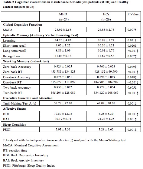
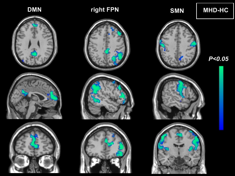
There were no significant differences in age, sex, or education level between MHD group and HCs (Table 1). Worse neuropsychological performances including MoCA, AVLT, n-back test, and TMT-A scores were found in MHD group compared with HCs (all P<0.05). The higher levels of depressed and anxious mood, the Sleep Condition were found in MHD group compared with HCs (all P<0.05) (Table 2). The 15 RSNs of interest in the study were, subsets of the large seven major RSNs identified by Yeo et al11: visual, somatomotor, dorsal attention, limbic, ventral attention, default, and frontoparietal networks. Compared with HCs, the MHD group showed decreased functional connectivity in the default mode network, the right frontoparietal network, and the sensorimotor network (P<0.05, FDR corrected) (Figure 2). No differences were observed for the dorsal attention network, ventral attention network, visual network, and limbic network. The decreased functional connectivity of the anterior cingulate cortex in the default mode network had a positive correlation with the MoCA scores. The decreased functional connectivity of the right inferior parietal lobule in the right frontoparietal network had a negative correlation with the TMT-A scores. The decreased functional connectivity of the right dorsal lateral prefrontal cortex had a negative correlation with the two-back reaction time scores in n-back working memory battery (P<0.05, FDR corrected).Discussion
The decrease functional connectivity of some critical brain network reflects cognitive deficit in brain function. In ESRD patients with maintenance hemodialysis, three resting-state subnetworks (DMN, SMN, and rFPN) showed decreased functional activity, four subnetworks (DAN, VAN, VN, and LN) remained unchanged. This suggests that ESRD patients had selective impairment instead of far-ranging impairment of resting-state subnetworks. As an important component of the DMN or the rFPN, the decreased functional connectivity of the PCC, as well as the DLPFC and IPL, are closely correlation with working-memory, execution function and attention in ESRD patients. Our results may contribute to understanding underlying neuropathological substrate of cognitive dysfunction in patients of ESRD.Conclusion
Our study demonstrates that ESRD patients with maintenance hemodialysis showed decreased functional connectivity in the default mode network, the right frontoparietal network, and the sensorimotor network, which have significant correlation with cognitive dysfunction. This study might contribute to better understanding the underlying neuropathological mechanism of cognitive dysfunction in patients with ESRD.Acknowledgements
This research was supported by the National Natural Science Foundation of China (Grant No. 81371530 and 81571640) and Natural Science Foundation of Shaanxi Province of China (Grant No. 2017ZDJC-13).References
- Foley RN, Collins AJ. End-stage renal disease in the United States: an update from the United States Renal Data System. J Am Soc Nephrol 2007; 18(10): 2644-8.
- Bugnicourt JM, Godefroy O, Chillon JM, Choukroun G, Massy ZA. Cognitive disorders and dementia in CKD: the neglected kidney-brain axis. Journal of the American Society of Nephrology: JASN. 2013; 24(3): 353-63.
- Agganis BT, Weiner DE, Giang LM, et al. Depression and cognitive function in maintenance hemodialysis patients. Am J Kidney Dis, 2010,56(4): 704-712.
- Ding D, Li P, Ma X Y, et al. The relationship between putamen-SMA functional connectivity and sensorimotor abnormality in ESRD patients. Brain Imaging & Behavior, 2017(5):1-9.
- Mu J, Chen T, Liu Q, et al. Abnormal interaction between cognitive control network and affective network in patients with end-stage renal disease. Brain Imaging & Behavior, 2018, 12(4): 1099-1111.
- Manjula Kurella Tamura, Kristine Yaffe. Dementia and cognitive impairment in ESRD: diagnostic and therapeutic strategies. Kidney International, 2011, 79(1): 14-22.
- Li P, Ding D, Ma X Y, et al. Altered intrinsic brain activity and memory performance improvement in patients with end-stage renal disease during a single dialysis session. Brain Imaging & Behavior, 2018(6): 1-10.
- Ni L, Wen J, Zhang L J, et al. Aberrant default-mode functional connectivity in patients with end-stage renal disease: a resting-state functional MR imaging study. Radiology, 2014, 271(2): 543-52.
- Jenkinson M, Beckmann C F, Behrens T E J, et al. FSL.Neuroimage 2012, 62(2): 782-90.
- Beckmann C F, Smith S M. Probabilistic independent component analysis for functional magnetic resonance imaging. IEEE Trans Med Imaging, 2004, 23(2): 137-152.
- B. T. Thomas Yeo, Fenna M. Krienen, Jorge Sepulcre, et al. The organization of the human cerebral cortex estimated by intrinsic functional connectivity. Journal of Neurophysiology, 2011, 106(3): 1125-1165.
Figures