3287
Altered topological organization in white matter connectome in Parkinson’s disease with and without rapid eye movement sleep behavior disorder1Second Affiliated Hospital of Zhejiang University School of Medicine, Hangzhou, China
Synopsis
We detected the alteration of white matter connectome in Parkinson’s disease (PD) patients with and without Rapid eye movement sleep behavior disorder (RBD). 155 PD patients including 66 possible RBD (pRBD) and 89 non-possible RBD (npRBD) and 71 normal controls were included. Diffusion-tensor magnetic resonance imaging and graph theory were used to explore the topologic organization of the brain structural connectome. Significant decreased nodal efficiency were found in specific regions including hippocampus and inferior occipital gyrus in PD-pRBD patients. Both these nodal properties were negatively correlated with RBD severity. This study may contribute to understand the pathophysiology of PD-RBD.
Introduction
Rapid eye movement (REM) sleep behavior disorder (RBD) is a common parasomnia characterized by loss of normal skeletal muscle atonia during REM sleep with prominent dreaming and motor activity1, 2. Studies show that it has a strong association with α synucleinpathies such as Parkinson’s disease (PD) 3, 4, where it can either occur during the course of the disease or appear at the prodromal phase of PD5. PD patients with RBD tend to have a poorer prognosis in terms of postural instability, gait disturbance and cognitive impairment compared with PD patients without RBD6, 7. Therefore, it is necessary to investigate the pathophysiological mechanism of RBD in PD patients, which may provide new insights to understand the substrate of PD. In the current study, we sought to investigate the altered topological properties of white matter connectome in PD patients with possible RBD (PD-pRBD) and non-possible RBD (PD-npRBD). We hypothesized that PD-pRBD may exhibited a disrupted brain network.Methods
155 PD patients including 66 possible RBD (pRBD) and 89 non-possible RBD (npRBD) and 71 normal controls (NCs) were included in the present study. Diffusion-tensor magnetic resonance imaging and graph theory were used to explore the topological organization of the brain network. Individual white matter (WM) networks were constructed with 116 nodes in whole brain including cerebellum and with considering the fiber number (FN) and fractional anisotropy (FA) as the weight of each edge. We analyzed small-worldness (σ), global and regional properties of WM connectome of three groups, and the network properties among groups were compared with a linear regression model.Results
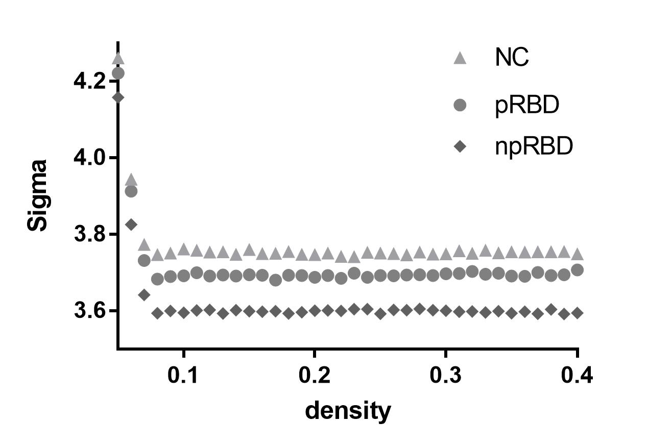
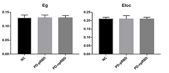
Inter-group comparisons of small-worldness and global network measures
We found that over the density range of 0.05 to 0.4, sigma was consistently greater than 1 for white matter connectome of NCs, PD-npRBD group and PD-pRBD group. Besides, there was no difference in AUC analyses of small-worldness and global network measures among three groups.
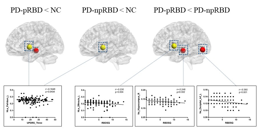
Inter-group comparisons of regional network measures
In comparisons with NCs, PD-pRBD exhibited decreased nodal efficiency in hippocampus, and both PD-pRBD and PD-npRBD showed lower nodal local efficiency in pallidum. When comparing with PD-npRBD, PD-pRBD showed lower nodal efficiency in hippocampus and inferior occipital gyrus, and lower nodal local efficiency in insula.
Correlations between disrupted nodal properties and clinical severity
We found that disrupted nodal efficiency was negatively correlated with RBD scores and decreased nodal local efficiency in insula was correlated with higher RBD scores in PD group. Furthermore, we found that decreased nodal local efficiency in pallidum was negatively associated with UPDRS Ⅲ scores in all PD patients.
Discussion
In this study, we applied graph theoretical analyses to compare white matter connectome of two PD groups (PD-pRBD and PD-npRBD) and controls, which would be helpful to make out the potential neural substrates of PD-pRBD. We found both PD-pRBD group, PD-npRBD group and controls group exhibited typical features of small-worldness, and there was no difference between any two groups in global network measures, which indicated that both two PD groups still keep a relatively integrated global function including abilities for information processing within and across anatomically interconnected brain regions. The reason for the reserved small world architectures maybe that our PD patients were at a relatively early stage, the mean Hoehn and Yahr stage was 1.6.
One of the main intriguing findings in our study was decreased nodal efficiency in hippocampus and inferior occipital gyrus as well as lower nodal local efficiency in insula in PD-pRBD when comparing with PD-npRBD. Both these properties were negatively correlated with RBD score. Several studies exhibited decreased gray matter volume of the hippocampus and impaired brain function as well as reduced cerebral blood flow in occipital areas8-10. Our results of decreased nodal properties in hippocampus and inferior occipital gyrus were consistent with these previous studies. Furthermore, the decreased nodal properties were negatively correlated with higher RBD severity, which indicated the specific regions were related to RBD symptoms in PD patients.
In addition, we found both PD-pRBD and PD-npRBD exhibited decreased nodal local efficiency in pallidum when comparing with NCs. Furthermore, decreased nodal local efficiency in pallidum was negatively correlated with UPDRS Ⅲ scores, which indicated that disrupt pallidum play a role in the impairment of motor function in PD patients. Disrupted function of basal ganglia has been found in several studies, and pallidum, a core region in basal ganglia, also exhibited impairment in PD patients11, 12.
Conclusion
Preserved global network properties indicated that early stage PD still keep a relatively integrated global function including abilities for information processing within and across anatomically interconnected brain regions. While disrupted nodal efficiency of the brain’s structural connectome in PD-pRBD provided novel information concerning the structural substrates of PD-pRBD.Acknowledgements
Tis work was supported by the 13th Five-year Plan for NationalKey Research and Development Program of China (No.2016YFC1306600). The authors acknowledge the whole staff of the Parkinson’sProgression Markers Initiative (PPMI) database.References
1. Olson EJ, Boeve BF, Silber MH. Rapid eye movement sleep behaviour disorder: demographic, clinical and laboratory findings in 93 cases. Brain 2000;123 ( Pt 2):331-339.
2. Schenck CH, Mahowald MW. REM sleep behavior disorder: clinical, developmental, and neuroscience perspectives 16 years after its formal identification in SLEEP. Sleep 2002;25(2):120-138.
3. Gagnon JF, Bedard MA, Fantini ML, et al. REM sleep behavior disorder and REM sleep without atonia in Parkinson's disease. Neurology 2002;59(4):585-589.
4. Sudarsky L, Friedman J. REM sleep behavior disorder: a possible early marker for synucleinopathies. Neurology 2006;67(11):2090; author reply 2090-2091.
5. Iranzo A, Molinuevo JL, Santamaria J, et al. Rapid-eye-movement sleep behaviour disorder as an early marker for a neurodegenerative disorder: a descriptive study. Lancet Neurol 2006;5(7):572-577.
6. Sinforiani E, Pacchetti C, Zangaglia R, Pasotti C, Manni R, Nappi G. REM behavior disorder, hallucinations and cognitive impairment in Parkinson's disease: a two-year follow up. Mov Disord 2008;23(10):1441-1445.
7. Postuma RB, Bertrand JA, Montplaisir J, et al. Rapid eye movement sleep behavior disorder and risk of dementia in Parkinson's disease: a prospective study. Mov Disord 2012;27(6):720-726.
8. Lim JS, Shin SA, Lee JY, Nam H, Lee JY, Kim YK. Neural substrates of rapid eye movement sleep behavior disorder in Parkinson's disease. Parkinsonism Relat Disord 2016;23:31-36.
9. Rahayel S, Montplaisir J, Monchi O, et al. Patterns of cortical thinning in idiopathic rapid eye movement sleep behavior disorder. Mov Disord 2015;30(5):680-687.
10. Vendette M, Gagnon JF, Soucy JP, et al. Brain perfusion and markers of neurodegeneration in rapid eye movement sleep behavior disorder. Mov Disord 2011;26(9):1717-1724.
11. Guan X, Zeng Q, Guo T, et al. Disrupted Functional Connectivity of Basal Ganglia across Tremor-Dominant and Akinetic/Rigid-Dominant Parkinson's Disease. Front Aging Neurosci 2017;9:360.
12. Szewczyk-Krolikowski K, Menke RA, Rolinski M, et al. Functional connectivity in the basal ganglia network differentiates PD patients from controls. Neurology 2014;83(3):208-214.
Figures