3285
The temporo-insular projection system: a multisubject fiber tractography study using connectome diffusion data1Department of Imaging and pathology, Translational MRI, KU Leuven, Leuven, Belgium, 2Department of Neurosciences, Research group experimental neurosurgery and neuroanatomy, KU Leuven, Leuven, Belgium
Synopsis
The precise structural connections between the amygdala, anterior temporal pole and insula remain poorly understood. These connections have been described with ex-vivo dissection, however the diffusion-based counterparts to these bundles haven’t been described in detail. In a recent study (currently under review) we investigated these connections with ex-vivo dissection on 11 brain specimens. We also explored the possibility of reconstructing the bundles found on dissection with Diffusion MRI. Here we demonstrate the results obtained for reconstructing these white matter connections using 20 randomly selected healthy participants of the HCP young adults data release.
INTRODUCTION
The structure of white matter fibers connecting the amygdala, anterior temporal pole and the insular cortex is poorly studied. These white matter tracts have important clinical relevance due to their role in propagation of epileptic activity. Ex-vivo, Klingler and Gloor described the ‘fasciculus amygdaloinsularis’1 & recent studies investigated these connections in primates2.
However, it has not been specifically reported in-vivo in humans, this may be due to the abundance of fiber crossings within these regions and the small size of the involved structures and bundles, which poses a technical problem for typical diffusion tensor imaging (DTI) methods3.
Here we investigate the diffusion correlate to these white matter connections found on virtual dissection using an advanced preprocessing pipeline on high spatial and angular resolution diffusion data of normal volunteers (n=20) acquired from the Human Connectome Project young adults preprocessed data release (v3.19.0 – released 1/3/2017).
METHODS
We used minimally preprocessed structural and diffusion images4 of the HCP young adults database acquired by the Washington University and University of Minnesota consortium5. Participants ages ranged between 22 – 35 years, demographics available at (https://db.humanconnectome.org/), 20 subjects were randomly selected as a preliminary sample.
Images were acquired on a Siemens 3T Skyra MRI scanner as previously detailed6. All images were preprocessed using the pipeline described by Glasser et al4, resulting in subject specific whole brain parcellation of the structural images using Freesurfer7, as well as diffusion weighted images which were denoised and corrected for gradient distortions as well as subject motion, eddy current artefacts and Echo planar imaging (EPI) distortions.
Each subject’s preprocessed T1, T2 weighted images and Freesurfer labels were warped to native diffusion space using ANTs8 and 5ttgen of Mrtrix39 was used to generate tissue probability maps. The diffusion data was processed using Mrtrix3. First, the preprocessed diffusion data was bias corrected using dwibiascorrect with ANTs N4 bias field correction10, followed by dwi2response and dwi2fod using the multi-shell multi-tissue framework11 to estimate tissue specific response functions and fiber orientation distributions (FOD). These FODs were then normalized for multiple tissues with mtnormalise. The individual T1s and corrected FODs were then used to generate population templates from the 20 participants with the population_template script of Mrtrix3. Whole brain tractography was done with FACT12 and with iFOD213, with anatomical constraint14 and dynamic seeding15 in both cases. We used tckedit to segment the whole brain tractograms using VOIs derived from the AAL atlas looking for streamlines connecting the insula, amygdala, and anterior temporal pole, constituting the amygdalo-insular (AIF) and temporo-insular (TIF) fiber bundles, respectively. Other VOIs (frontal and occipital) were used for the Uncinate and Inferior Fronto-occpital fasciculus (UF and IFOF).
RESULTS
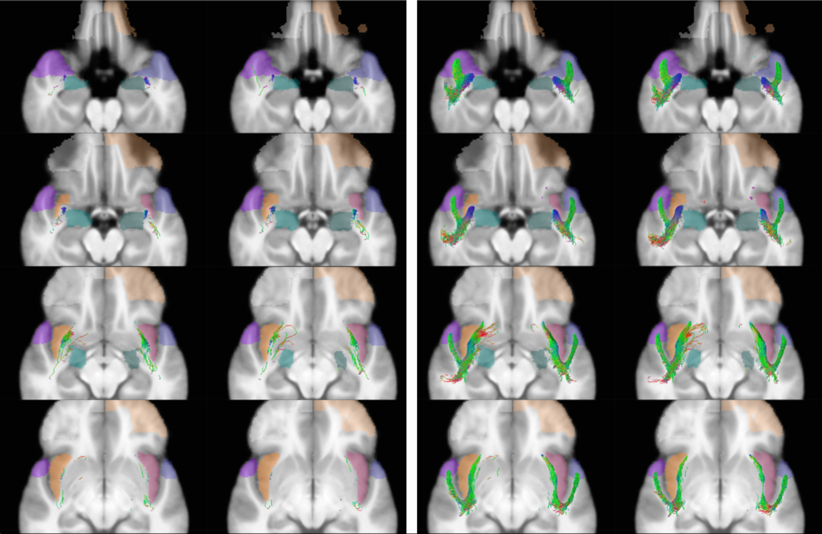
FACT tractography only gives a plausible result for the TIF, with considerable underestimation of its volume and extent, whereas it fails to depict a plausible result for the AIF. CSD however, demonstrates both fiber bundles (Figure 1). The AIF is noted to extend from the antero-ventro-lateral aspect of the amygdala to the anterior insula. We also found the TIF to be projecting from the anterior temporal pole to the insula, with a fanning out of its fibers at the level of the extreme capsule, ascending toward the different insular gyri.DISCUSSION
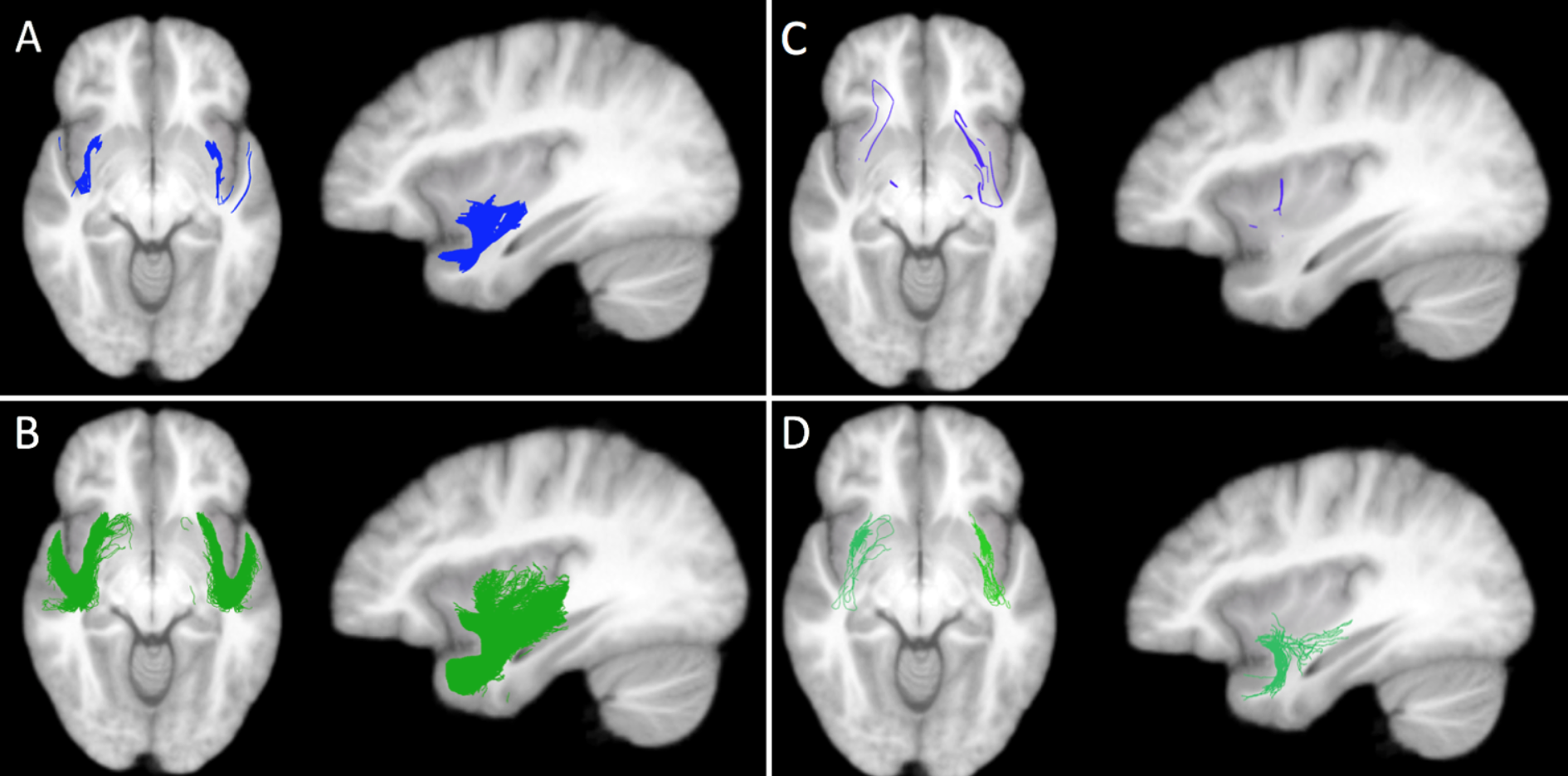
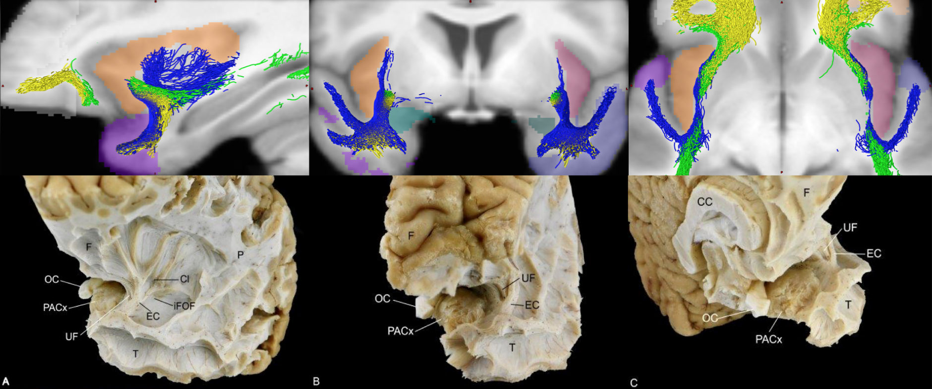
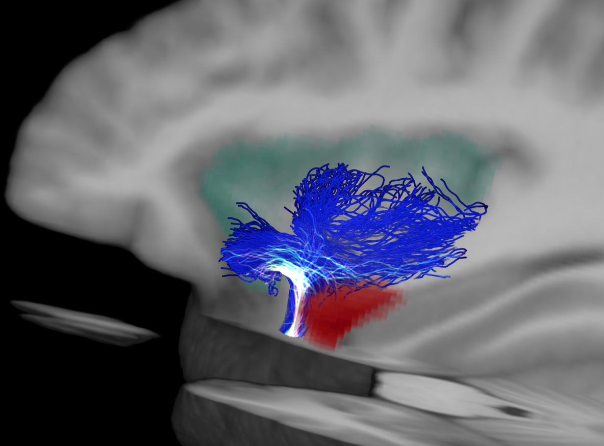
The failure of FACT to reconstruct the AIF was expected, due to the abundance of crossing fibers in the temporal stem, which is a known weakness of FACT3. CSD demonstrated both fiber bundles within the temporal short association fibers (Figure 2). To further investigate the structure and anatomical relations of these bundles we tracked the UF and the IFOF. Both the UF and IFOF fibers run within the temporal stem. We found these bundles being more medial, while the AIF and TIF occupy a more lateral location. These findings are consistent with ex-vivo dissection16, localizing the AIF and TIF to the extreme capsule, and the UF and IFOF to the external capsule (Figure 3). Furthermore, our results seem to indicate that the AIF is a subset of the TIF (Figure 4).CONCLUSION AND FUTURE DIRECTIONS
We found imaging evidence supporting the existence of the AIF and TIF, while shedding light on the role of probabilistic CSD in investigating some of the yet poorly understood white matter connections, especially those that reside in areas with an abundance of crossing fibers. We plan to expand this work to include 100 healthy subjects from the HCP database to investigate the reproducibility of our results and to investigate inter-individual variability of these bundles.Acknowledgements
No acknowledgement found.References
1. Klingler J, Gloor P. The connections of the amygdala and of the anterior temporal cortex in the human brain. J Comp Neurol. 1960;115(3):333-369.
2. Mufson EJ, Mesulam M-M, Pandya DN. Insular interconnections with the amygdala in the rhesus monkey. Neuroscience. 1981;6(7):1231-1248.
3. Lee D-H, Park JW, Park S-H, Hong C. Have You Ever Seen the Impact of Crossing Fiber in DTI?: Demonstration of the Corticospinal Tract Pathway. PLoS One. 2015;10(7):e0112045.
4. Glasser MF, Sotiropoulos SN, Wilsonc JA, et al. The Minimal Preprocessing Pipelines for the Human Connectome Project. Neuroimage. 2013;October(15):105-124.
5. Van Essen DC, Smith SM, Barch DM, et al. The WU-Minn Human Connectome Project: an overview. Neuroimage. 2013;80:62-79.
6. WU-Minn HCP 1200 Subjects Data Release Reference Manual.; 2017.
7. Fischl B. FreeSurfer. Neuroimage. 2012;62(2):774-781.
8. Avants BB, Tustison NJ, Song G, et al. A Reproducible Evaluation of ANTs Similarity Metric Performance in Brain Image Registration. Neuroimage. 2011;54(3):2033-2044.
9. Tournier JD, Calamante F, Connelly A. MRtrix: Diffusion tractography in crossing fiber regions. Int J Imaging Syst Technol. 2012.
10. Tustison NJ, Avants BB, Cook PA, et al. N4ITK: Improved N3 Bias Correction. IEEE Trans Med Imaging. 2010;29(6):1310-1320.
11. Jeurissen B, Tournier J-D, Dhollander T, Connelly A, Sijbers J. Multi-tissue constrained spherical deconvolution for improved analysis of multi-shell diffusion MRI data. Neuroimage. 2014;103:411-426.
12. Mori S, Kaufmann WE, Davatzikos C, et al. Imaging cortical association tracts in the human brain using diffusion-tensor-based axonal tracking. Magn Reson Med. 47(2):215-223.
13. Tournier J-D, Calamante F, Connelly A. Improved probabilistic streamlines tractography by 2nd order integration over fibre orientation distributions. Proc Intl Soc Mag Reson Med. 2010;18.
14. Smith RE, Tournier J-D, Calamante F, Connelly A. Anatomically-constrained tractography: Improved diffusion MRI streamlines tractography through effective use of anatomical information. Neuroimage. 2012;62(3):1924-1938.
15. Smith RE, Tournier J-D, Calamante F, Connelly A. SIFT2: Enabling dense quantitative assessment of brain white matter connectivity using streamlines tractography. Neuroimage. 2015;119:338-351.
16. Nachtergaele P, Radwan A, Swinnen S, et al. The temporoinsular projection system: an anatomical study. J Neurosurg (Under review).
Figures