3283
Neural mechanism of processing speed decline induced by anatomically connected white matter tract damage and cortical function abnormality1Department of radiology, 2nd affiliated hospital of Zhejiang university school of medicine, Hangzhou, China, 2GE Healthcare, Shanghai, China
Synopsis
White matter hyper-intensities (WMH) is considered as an important source of morbidity associated with dementia, stroke and increased mortality risk. Previous studies have suggested that WMH always leads to the decline of information processing speed, which have an impact on activities of daily living. Current hypothesis is that the dysfunctions caused by WMH is the result of “disconnection”, while the connection between the structural and functional alteration has not been fully investigated. We aimed to explore the underlying pathway of information processing speed decline via combining the spatial distribution of WMH, microstructural changes based tractography and cortical activity alterations. The results from different modalities converged in the occipital lobe with precise spatial overlappings. Results show regional WMH may indicate disrupted tract integrity and cause altered brain activities, leading to impaired function. This WMH-tract-function-behavior link is critical for WMH induced dysfunctions and treatment strategies.
Introdunction
White matter hyper-intensities (WMH), a common sign in elder population, appeared as hyper-intensity patches in the white matter on T2 and T2FLAIR image1-2. WMH is considered as an important source of morbidity associated with dementia, stroke and increased mortality risk3-5. Previous studies suggested that WMH always leads to executive dysfunctions, especially the decline of information processing speed, which have an impact on activities of daily living6-8. There are several ways to explore the underlying pathway of dysfunction caused by WMH - for example analyzing the distribution of WMH lesion and the integrity of specific white matter tracts9-18, or assessing the connectivity of brain networks11,19. While most previous studies only analyzed the relationship between information processing ability and only one or two of the three above-mentioned aspects, without fully exploring the WMH-Tract-Function-Behavior link13,16-17,20-23. In order to better understand the underlying pathway between the anatomical and spatial proximity of WMH lesion, white matter tract damage and cortical damage related with a specific domain, we aimed to investigate how WMH leads to information processing dysfuntion through a comprehensive assessment of brain features.Methods
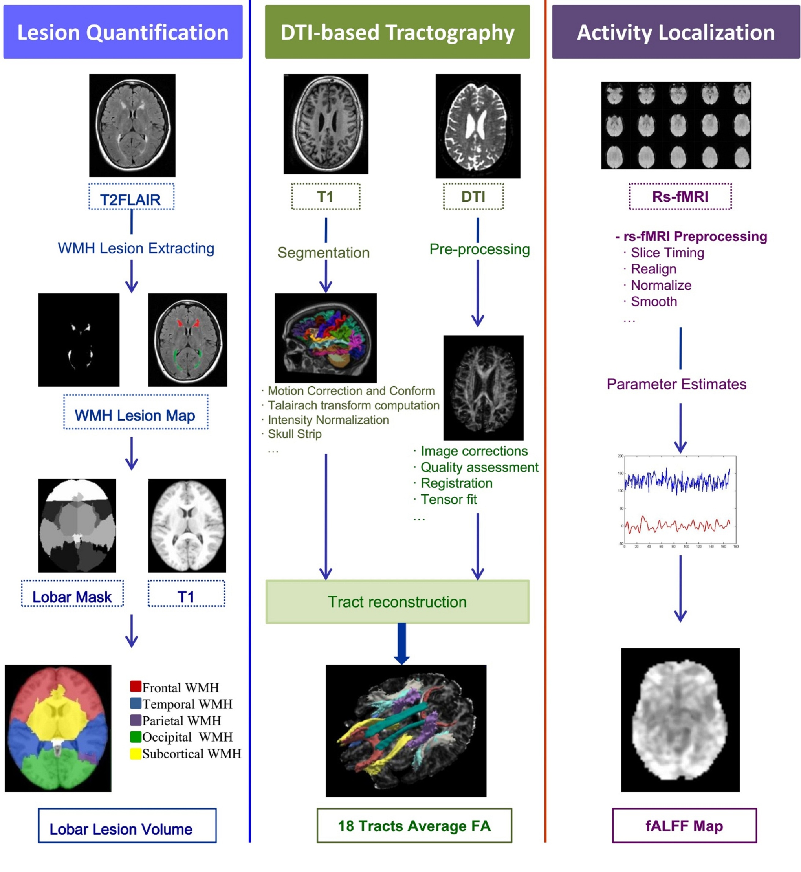
We included sixty elder adults. Information processing ability was assessed by the trail making test part A (TMT-A), and multi-modal magnetic resonance images were acquired. Workflow of multi-modalities imaging pre-processing was presented in Fig. 1. For analyzing the distribution of WMH volumes, we extracted WMH volumes in frontal, occipital, temporal, parietal lobe and suncortical region based on T2FLAIR. TRACULA (TRActs Constrained by UnderLying Anatomy)24, a probabilistic tractographic method was used to obtain the mean fractional anisotropy (FA) values of 18 major tracts. Whereas voxel-wise global analysis of cortical function was estimated by the fractional Amplitude of Low-Frequency Fluctuations (fALFF) value based on fMRI. Correlation analysis was performed to assess the relationship between TMT-A and several brain features, including WMH volumes in each brain lobe, diffusivity parameters in major white matter tracts, and regional brain activities across the whole brain. Finally, a multiple linear regression analysis was used to demonstrate the contribution of each index to information processing speed alterations.Results
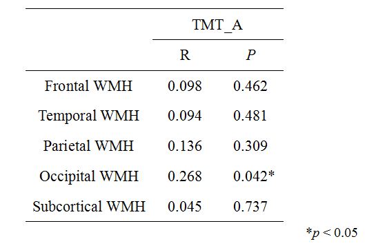
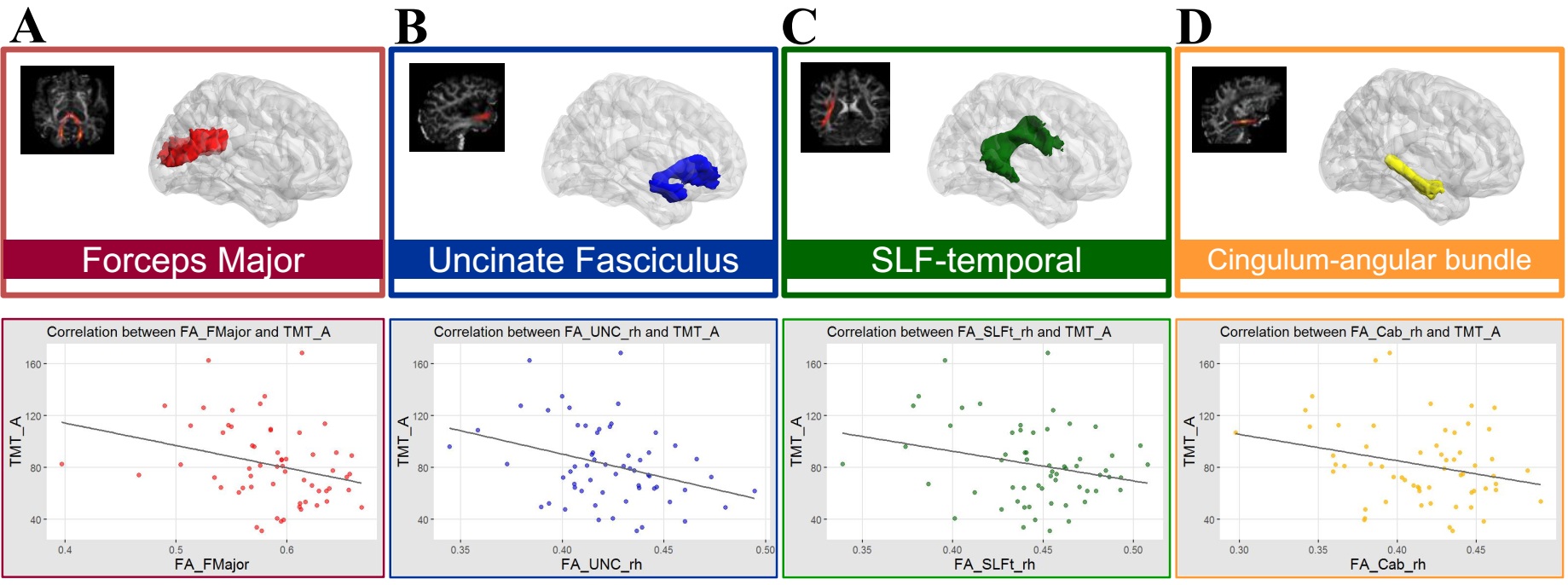
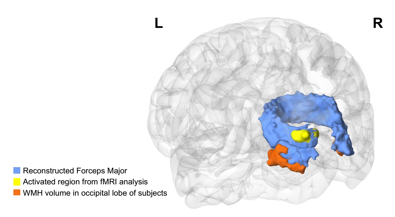
We found that the WMH volume in occipital lobe(Table 1) and FA of forceps major, an occipital association fiber, were significantly correlated with TMT-A scores(Fig. 2). Besides, resting-state brain activities in the visual association cortex connected to the forceps major were also correlated with TMT-A scores(Fig. 3). Multiple linear regression showed that FA of forceps major and regional brain activities were significant predictors of TMT-A scores.Discussion
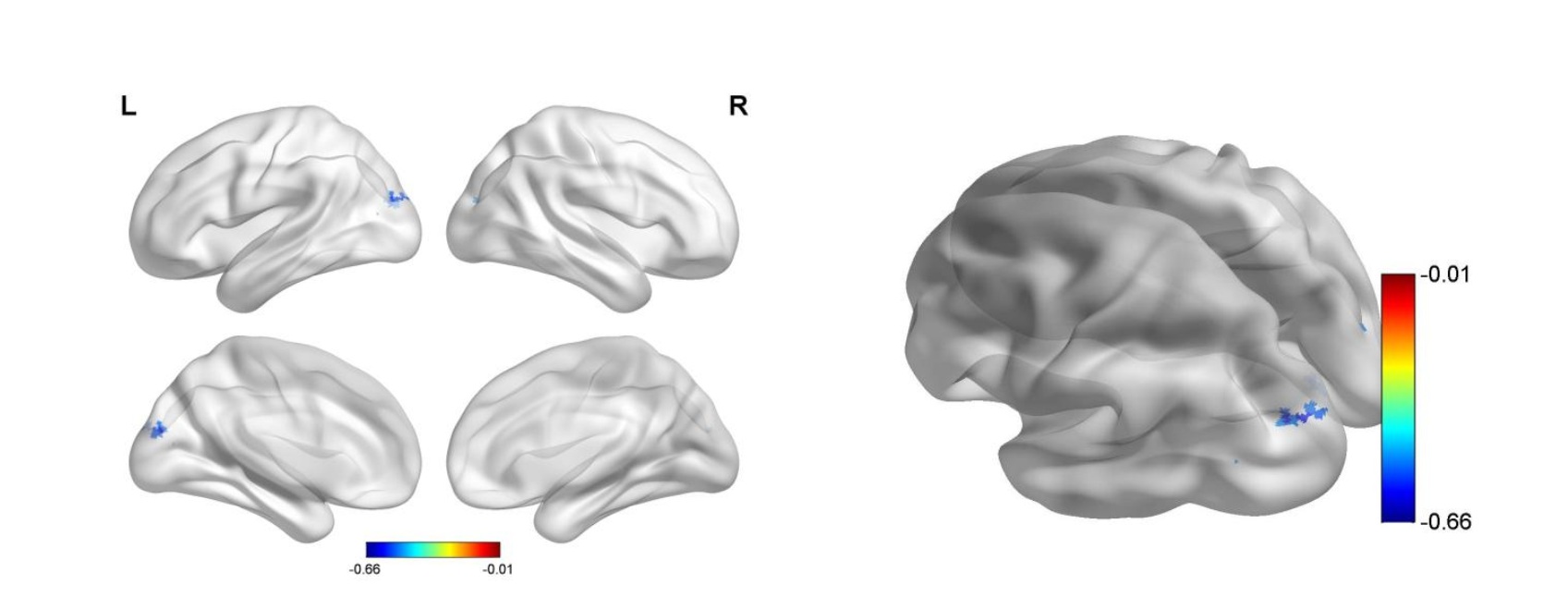
In this study, we performed a combined analysis of macrostructure, microstructure, and cortical function assessment to explore the mechanism of information processing speed changes induced by WMH. Interestingly, results from the three modalities converged in the occipital lobe, with precise spatial overlappings in visual association cortex (Fig. 4). Besides, multiple regression analysis revealed that FA in FMajor and local fALFF could predict the TMT-A score. We suggest that in subjects with WMH, the poorer TMT performance was largely due to damaged visual-spatial information processing abilities. Furthermore, we identified consistent alterations in the occipital lobe. We suggest that the WMH in occipital lobe might lead to the impairment of the forceps major integrity, and the neural circuit between the visual association cortex connected by WM tracts may be broken. That caused a decline in visual information conduction efficiency, which ultimately resulted in the alteration of visuospatial information processing ability. These findings provided strong support for the the WMH-Tract-Function-Behavior theoretical framework about the WMH-mediated dysfunction.In summary, the underlying pathway of information processing dysfunction caused by WMH may involve in multi-domain, but the alteration of visuospatial information processing ability is remarkable in our WMH population. The degradation of the WM microstructures and neural disconnection may further disrupt the parietal-occipital network and frontal-occipital network. Our results indicated that the processing of WMH-mediated changes in information processing ability could have a strategic mechanism, which may implicate a structural-functional pathway highly corresponding in anatomical structure and function. However, future studies are needed to confirm such a hypothesis.Conclusion
In conclusion, this study explored the underlying mechanism of WMH-related information processing speed deline using a multi-modality approach. A variety of structural and functional alterations were found related to information processing ability, and results from different modalities converged in the occipital lobe, highlighting that visuospatial dysfunction is important for trail-making speed. Besides, the precise spatial overlapping of lesion-tract-function results provided a unique opportunity for understanding how WMH pathologies lead to brain dysfunction. Given the link between WMH and neurovascular disease, exploring the neural mechanism of information processing speed changes induced by WMH could also provide evidence on preventive and treatment strategies.Acknowledgements
No acknowledgement found.References
1. Wardlaw, J.M., et al., Neuroimaging standards for research into small vessel disease and its contribution to ageing and neurodegeneration. Lancet Neurol, 2013. 12(8): p. 822-38.
2. Fazekas, F., et al., Pathologic correlates of incidental MRI white matter signal hyperintensities. Neurology, 1993.43(9): p. 1683-1683.
3. Stefaniak, J.D., et al., Enzyme replacement therapy and white matter hyperintensity progression in Fabry disease. Neurology, 2018. 91(15): p. e1413-e1422.
4. Brickman, A.M., et al., Reconsidering harbingers of dementia: progression of parietal lobe white matter hyperintensities predicts Alzheimer's disease incidence. Neurobiology of Aging, 2015. 36(1): p.27-32.
5. Pantoni, L., Cerebral small vessel disease: from pathogenesis and clinical characteristics to therapeutic challenges. Lancet Neurol, 2010. 9(7): p. 689-701.
6. Prins, N.D., et al., Cerebral small-vessel disease and decline in information processing speed, executive function and memory. Brain, 2005. 128(9): p. 2034-2041.
7. Debette, S., et al., Association of MRI Markers of Vascular Brain Injury With Incident Stroke, Mild Cognitive Impairment, Dementia, and Mortality. Stroke, 2010. 41(4): p. 600-606.
8. Kloppenborg, R.P., et al., Presence and progression of white matter hyperintensities and cognition A meta-analysis. Neurology, 2014. 82(23): p. 2127-2138.
9. Sun, J., et al., The relationship between microvasculature in white matter hyperintensities and cognitive function. Brain Imaging and Behavior, 2017. 11(2): p. 503-511.
10. Smith, E.E., et al., Correlations between MRI white matter lesion location and executive function and episodic memory. Neurology, 2011. 76(17): p. 1492-9.
11. Sudo, F.K., et al., Dysexecutive syndrome and cerebrovascular disease in non-amnestic mild cognitive impairment: a systematic review of the literature. Dementia & Neuropsychologia, 2012. 6(3): p. 145-151.
12. Bolandzadeh, N., et al., Pathways linking regional hyperintensities in the brain and slower gait. NeuroImage, 2014. 99: p. 7-13.
13. Lampe, L., et al., Lesion location matters: The relationships between white matter hyperintensities on cognition in the healthy elderly. Journal of Cerebral Blood Flow & Metabolism, 2017: p. 0271678X1774050.
14. Papma, J.M., et al., Cerebral small vessel disease affects white matter microstructure in mild cognitive impairment. Human Brain Mapping, 2014. 35(6): p. 2836-2851.
15. Ghanavati, T., et al., Deep white matter hyperintensities, microstructural integrity and dual task walking in older people. Brain Imaging and Behavior, 2018.
16. Johnson, N.F., et al., Endothelial Function Is Associated with White Matter Microstructure and Executive Function in Older Adults. Frontiers in Aging Neuroscience, 2017. 9.
17. Tuladhar, A.M., et al., White matter integrity in small vessel disease is related to cognition. NeuroImage: Clinical, 2015. 7: p. 518-524.
18. Duering, M., et al., Strategic role of frontal white matter tracts in vascular cognitive impairment: a voxel-based lesion-symptom mapping study in CADASIL. Brain, 2011. 134(8): p. 2366-2375.
19. Jacobs, H.I.L., et al., The association between white matter hyperintensities and executive decline in mild cognitive impairment is network dependent. Neurobiology of Aging, 2012. 33(1): p. 201.e1-201.e8.
20. Hirsiger, S., et al., Executive Functions in Healthy Older Adults Are Differentially Related to Macro- and Microstructural White Matter Characteristics of the Cerebral Lobes. Frontiers in Aging Neuroscience, 2017. 9.
21. Tullberg, M., et al., White matter lesions impair frontal lobe function regardless of their location. Neurology, 2004.63(2): p. 246 - 253.
22. Smith, E.E., et al., Correlations between MRI white matter lesion location and executive function and episodic memory. Neurology, 2011. 76(17): p. 1492-9.
23. Zheng, J.J.J., et al., Brain White Matter Hyperintensities, Executive Dysfunction, Instability, and Falls in Older People: A Prospective Cohort Study. The Journals of Gerontology Series A: Biological Sciences and Medical Sciences, 2012. 67(10): p. 1085-1091.
24. Yendiki, A., Automated probabilistic reconstruction of white-matter pathways in health and disease using an atlas of the underlying anatomy. Frontiers in Neuroinformatics, 2011. 5.
Figures