3279
The information flow pattern of the human brain based on high temporal resolution functional MRI1Clinical Science, Philips Healthcare, Beijing, China, 2Capital Medical University, Beijing, China
Synopsis
To investigate the information flow pattern of human brain by using high temporal resolution functional MRI based on linear, non-linear Granger causality analysis and transfer entropy
Purpose
To investigate the information flow pattern of human brain by using high temporal resolution functional MRI.Materials and methods
It is an initial study, so only seven (Female/Male: 4/3, Age=35y with range of 27y-43y) high temporal resolution fMRI images (TR=0.72s with Simultaneous Multi-Slice Excitation and Acquisition and totally 1200 time points) download from Human Connectome Project (HCP) were included in this study. All the images were already processed according to HCP processing pipeline, in which some noise (signal drift, motion information, physiological noise and other signals which looks unlike brain BOLD signals) were excluded from the fMRI time series based on Independent Component Analysis (ICA) except for white and CSF signal regression, signal filtering, and image smoothing. In our following processing, we carried out the white and CSF signal regression, signal filtering with bandwidth of 0.001-0.1Hz limited to the grey matter region but no image smoothing to avoid the loss of images details due to smoothing. Linear and two kinds of non-linear Granger Causality Analysis (GCA) as well as transfer entropy (TE) were adopted to represent the causality or exchanged information between each pair of brain regions (defined by AAL 90 atlas). We did not adopt the absolute value of the GCA or TE but the relative value (which we called directed information flow) between each pair of brain regions (which was also called nodes in functional network).
Taking TE for example, for two node i and node j, the directed information flow (here DTE) between them was defied as
DTE(i)=TE(i,j)/(TE(i,j)+TE(j,i))
For node with DTE>0.5, this node is defined as the driving node in an information exchange path, and for node with DTE>0.5, this node is defined as the receiving node in an information exchange path. For node with DTE=0.5, we think there is no directed information exchange between nodes.
For the whole brain information flow pattern analysis, the mean DTE values of the node as driving and receiving information were calculated respectively and thus each brain node were given a mean DTE for its function of driving (mean DTE>0.5 as driving information) or receiving (mean DTE>0.5 as receiving information). The above operations were also applied for linear and two types of non-linear GCA for the directed information flow representation.
Results
The information flow patterns were showed in Figure 1. The results of all the four methods showed that the information flow of human brain were mainly from brain anterior area to posterior area. The results of GCA with different algorithms were similar. And it was also similar to the results of TE. Most parts of the frontal and temporal areas act as the driving node but most parts of the parietal and occipital act as the receiving nodes. Another finding is that the driving areas especially for frontal areas are located at the left hemisphere.Discussion
Granger causality (GC) used predictability to describe the causation between time series[1, 2]. The Granger cause of variable X to Y was defined when the predictability of Y by Y’s history information was decreased when the history information of X was removed. GC was firstly proposed to solve the causality in linear system. And then it was extend to nonlinear coupling. Transfer entropy (TE) is a variation of GC, which was based on information theory to infer the prediction ability of variable X for Y. That is equal to the reduction of prediction uncertainty of Y by both X and Y history information compared to only by Y’s history information. TE was suitable for system with large sample points such as EEG and MEG signals. Conventional fMRI only have very limited sample points (e.g. 100-300) which usually cause the bias of the probability estimation. Functional MRI in HCP have 1200 time points for 3T MR and 900 time points for 7T MR. And TE might be used to calculate the prediction information of different brain regions based on the high-temporal resolution images. The results showed that the TE have similar ability as GC to infer the causality of different brain regions. And found the anterior-to-posterior information pattern in healthy brain. The results was similar to the previous work[3] which indicated the anterior-to-posterior information pattern by using MEG in brain low-frequency activities.Conclusion
Human brain information flow pattern might be described by using high temporal resolution fMRI, which would be helpful for the studies of neuroscience.Acknowledgements
No acknowledgement found.References
[1] N. Wiener, “The theory of prediction,” in Modern Mathematics for the Engineer, edited by E. F. Beckenbach (McGraw-Hill, New York, NY,1956), pp. 125–139.
[2] C. W. J. Granger, “Time series analysis, cointegration, and applications. Nobel Lecture, December 8, 2003,” in Les Prix Nobel. The Nobel Prizes 2003, edited by T. Frängsmyr (Nobel Foundation, Stockholm, 2004), pp. 360–366.
[3] H. Arjan et al., Direction of information flow in large-scale resting-state networks is frequency-dependent. PNAS. 2016, 113 (14) 3867-3872
Figures
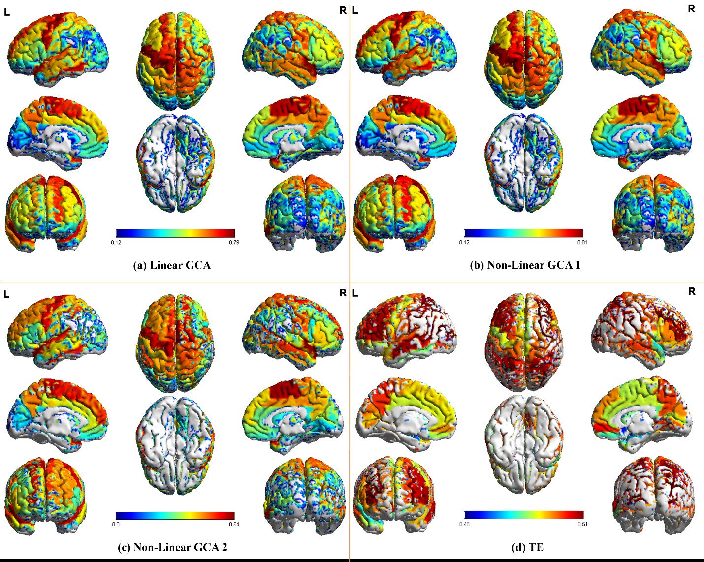
Figure 1.
The information flow pattern of human brain derived by linear GCA (a),
non-linear GCA 1 (b), non-linear GCA 2 (c) and TE (d).