3276
Longitudinal functional MRI for animal studies of neurovascular coupling in healthy ageing1Department of Neuroscience, Psychology and Behaviour, University of Leicester, Leicester, United Kingdom, 2School of Psychology, University of Nottingham, Nottingham, United Kingdom, 3Core Biotechnology Services, University of Leicester, Leicester, United Kingdom
Synopsis
Due to effects of anaesthetic on the BOLD signal, or use of toxic anaesthetics, longitudinal preclinical fMRI studies are uncommon, and studies of age-related disease progression show high variability and use large numbers of animals. This study uses a novel anaesthetic protocol in 11 rats to study changes in the BOLD signal with age. A strong, reproducible BOLD response to forepaw stimulation is found between 7 and 12 months old, and at 15 months old the number of active voxels is reduced by half. This shows that the protocol is suitable for longitudinal studies of ageing.
Introduction
Preclinical fMRI is a valuable tool in the understanding of disease mechanisms in animal models, and in preclinical drug development. The non-invasive nature of MRI can facilitate translation between studies on animal models and human subjects. However, preclinical fMRI does have its drawbacks. Animals require anaesthesia for scanning and commonly used anaesthetics such as isoflurane inhibit vascular reactivity. Anaesthetics without this effect such as urethane and alpha-chloralose are carcinogenic and so are restricted for terminal scans1. This has led to a lack of longitudinal preclinical fMRI studies, with those that do exist reporting inconsistent findings, due to the limitations of the anaesthetic protocol2. Also, no preclinical studies exist of changes in the haemodynamic response in normal ageing, which limits the application of fMRI in studying the progression of age-related disease. This study examines the suitability of using a novel, minimally invasive anaesthesia protocol in combination with a functional MRI protocol to assess alterations in neuronal activity due to physiological aging. Data is presented from the first four time points of an ongoing 18 month aging study in ratsMethods
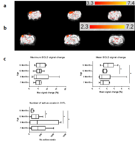
This study was conducted in accordance with the UK Animals (Scientific Procedures) Act, 1986 and following institutional ethical approval. 11 female Wistar Han rats aged 3 months were housed in standard cages and given daily access to a playpen to promote social and physical enrichment. Animals were scanned at 7, 9, 12 and months of age. Under 3% isoflurane anaesthesia, tail vein cannulation was performed before transferring the animals to the MRI bed. A bolus of 9mg/kg propofol3 was administered over 1 minute and isoflurane was gradually reduced to 0%. Respiration was monitored using a respiration pillow, temperature monitored using a rectal probe, and heart rate and blood oxygen saturation monitored using a pulse oximeter. Copper electrodes were inserted subcutaneously into the dorsal surface of the right forepaw between digits 1-2 and 2-3. 3 minutes after the bolus ended, a continuous infusion of propofol was given at 54mg/kg/hr for the duration of the scan. Rats were switched from breathing oxygen to room air and fMRI was performed for 9 minutes using a rapid EPI sequence (TR=250ms, TE=22ms, kzero=8, shots=2, data matrix =128x128). The forepaw was stimulated at 10mV, 10Hz, pulse width 1us, with a block design of 60s off, 30s on. A standard fMRI analysis pipeline was performed using FSL (www.fmrib.ox.ac.uk/fsl). Motion correction (MCFLIRT), brain extraction (rBET), bias field correction (FAST) and independent component analysis for artefact removal (MELODIC) were performed prior to time-series analysis in FEAT to visualise the BOLD response.Results
No significant difference in maximum BOLD signal change, mean BOLD signal change or number of active voxels was found between 7, 9 and 12 months. At 15 months, a significant decrease in number of active voxels (255+179 at 12 months to 114+45 at 15 months, P<0.05), and an increase in mean BOLD signal change (1.6+0.3% at 12 months to 2.1+0.8% at 15 months, P<0.05) were observed.Discussion
This study presents the first four time points of an 18 month ageing study in rats. The anaesthetic protocol showed no adverse effects from repeat imaging, a robust and reproducible BOLD response, with rapid recovery of the animals. Though some outliers were present at 9 months, the minimal change in signal between the first three time points supports the reproducibility of the protocol. The reduction in number of active voxels observed here is consistent with observations of age-related changes in humans4. The observed increase in BOLD signal amplitude is not consistent with human studies however5. Future work as the cohort ages further, including ongoing analysis to combine BOLD results with MRS and ASL, may help explain this finding. The consistency of results in younger rats followed by reduction in active voxels with age suggests that this model is suitable for longitudinal fMRI studies of ageing and disease progression.Acknowledgements
MRC IMPACT doctoral training partnership.
University of Leicester Biomedical Workshop.
University of Leicester Division of Biomedical Services animal technicians
References
1. Tremoleda et al (2012) Anaesthesia and physiological monitoring during in vivo imaging of laboratory rodents: considerations on experimental outcomes and animal welfare. EJNMMI Res 2(1):44.
2. Dijkhuizen et al (2012) Functional MRI and diffusion tensor imaging of brain reorganization after experimental stroke. Trans Stroke Res 3(1):36.
3. Griffin et al (2010) Propofol allows precise quantitative arterial spin labelling functional magnetic resonance imaging in the rat. NeuroImage 51(4):1395.
4. Huettel et al (2001) The effects of aging upon the hemodynamic response measured by functional MRI. NeuroImage 13: 161–175
5. Ances et al (2009) Effects of Aging on Cerebral Blood Flow, Oxygen Metabolism, and Blood Oxygenation Level Dependent Responses to Visual Stimulation. Hum Brain Mapp. 30(4): 1120–1132
Figures