3275
Longitudinal functional MRI mapping changes in neurovascular responses following middle cerebral artery occlusion1Department of Neuroscience, Psychology and Behaviour, University of Leicester, Leicester, United Kingdom, 2School of Psychology, University of Nottingham, Nottingham, United Kingdom, 3Core Biotechnology Services, University of Leicester, Leicester, United Kingdom
Synopsis
Due to limitations in anaesthetic protocol, fMRI studies of recovery following ischaemic stroke in rodents show high variability and must use large numbers of animals. This study uses a novel anaesthetic protocol which gives a highly reproducible BOLD signal in healthy animals to study changes in response to forepaw stimulation in 10 rats following middle cerebral artery occlusion. Even with the improved protocol, high variability between animals following MCAO is a confounding factor, and while a trend towards hyperactivation followed by return to baseline is seen, a larger number of animals is still required for longitudinal studies.
Introduction
Ischaemic stroke is one of the most common causes of death or disability. Survivors often develop motor deficits, and the effectiveness of physical therapy on motor recovery varies greatly between patients. Human fMRI studies have determined that recovery of motor function follows a two stage pattern of hyperactivation1 followed by focusing of activity as motor abilities are relearned. However, this pattern has not been observed in preclinical studies, due to the effects of previous anaesthetic protocols for longitudinal studies on the BOLD response2. To better understand post stroke recovery and develop treatments for deficits, preclinical models should demonstrate the same disease progression as humans. Here, a novel, minimally invasive anaesthesia protocol, was used to investigate longitudinal changes in the BOLD response in the primary somatosensory cortex (S1FL) following stroke.Methods
This study was conducted in accordance with the UK Animals (Scientific Procedures) Act, 1986 and following institutional ethical approval. 11 male Sprague-Dawley rats aged 3 months were housed in standard cages and given daily access to a playpen to promote social and physical enrichment. Animals were scanned one week prior to MCAO, and at 4 months of age, underwent 60 minute middle cerebral artery occlusion (MCAO) surgery to the right hemisphere. Animals were scanned at 48 hours, 1 week, 2 weeks, 3 weeks and 6 weeks following MCAO, and all animals were humanely killed following the 6 week scan. Animals were scanned under 54mg/kg.hr propofol anaesthesia3. fMRI was performed on three slices centred over bregma for 9 minutes using a rapid EPI sequence (TR=250ms, TE=22ms, kzero=8, shots=2, data matrix =128x128). The forepaw was stimulated at 10mV, 10Hz, pulse width 1us, with a block design of 60s off, 30s on Motion correction (MCFLIRT), brain extraction (rBET), bias field correction (FAST) and independent component analysis for artefact removal (MELODIC) were performed on fMRI data prior to time-series analysis in FEAT to visualise the BOLD response.Results
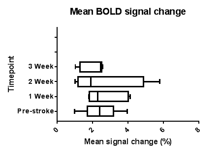
In all but one animal, the BOLD response in the injured hemisphere to contralesional forepaw stimulation was completely eliminated 2 days post MCAO, before recovering at later time points in most animals. Later time points showed no significant difference in BOLD response compared to the pre-MCAO time point. However, the standard deviation of both maximum BOLD signal and mean BOLD signal at 1, 2 and 3 weeks post-stroke was twice as large as the standard deviation at the pre-stroke time point. One animal showed no recovery over the 6 weeks. BOLD signal in two animals remained at baseline for all 6 weeks. In the remaining seven animals, when the BOLD signal returned, maximum and mean signal change was larger than at the pre-stroke time point, returning to baseline over subsequent weeks. The time at which the BOLD response returned in these animals varied between 1 week and 6 weeks.Discussion
While BOLD signal change following stroke was not significantly different from baseline, this can be attributed to the high variatiability between animals. Previous studies have shown that BOLD responses following stroke were highly variable between animals, and the time at which the BOLD response returned was also variable4. Our preliminary data in healthy ageing animals show that the anaesthesia and fMRI protocol gives a highly reproducible BOLD signal. Here, our data shows that, despite using a reproducible longitudinal fMRI method, there was still high variability in recovery between post-stroke rats. Thus, because of such high variability in the stroke model, previous studies using multiple groups or a less reproducible anaesthesia protocol may introduce additional confounding factors to the results. Comparison of pre-stroke s1FL activation with 1st post-stroke activation and 2nd post stroke activation showed a trend towards hyperactivation (max. BOLD signal changes: 5.097% pre-stroke, 8.57% post-stroke), however due to the low number of animals this was not statistically significant. Combination of this minimally invasive anaesthesia protocol and factoring in change in lesion volume over time into analysis may help minimise variability in the stroke model, and allow progression of stroke recovery to be studied using fewer animals.Acknowledgements
MRC IMPACT Doctoral training partnership
University of Leicester Biomedical Workshop
University of Leicester Division of Biomedical Services animal technicians
References
1. Tombari et al (2004) A longitudinal fMRI study: in recovering and then in clinically stable sub-cortical stroke patients. NeuroImage 23(3): 827
2. Dijkhuizen et al (2012) Functional MRI and diffusion tensor imaging of brain reorganization after experimental stroke. Trans Stroke Res 3(1):36.
3. Griffin et al (2010) ) Propofol allows precise quantitative arterial spin labelling functional magnetic resonance imaging in the rat. NeuroImage 51(4):1395. #
4. Weber et al (2008) Early prediction of functional recovery after experimental stroke: functional magnetic resonance imaging, electrophysiology, and behavioral testing in rats. J. Neurosci 28(5):1022–1029
Figures