3274
Altered resting-state functional connectivity of primary visual cortex in young patients with comitant exotropia1Radiology, Renmin Hospital of Wuhan University, Wuhan, China, 2Ophthalmology, Renmin Hospital of Wuhan Uiversity, Wuhan, China, 3GE Healthcare, Beijing, China, 4Radiology, Renmin Hospital of Wuhan Uiversity, Wuhan, China
Synopsis
The comitant exotropia (CE) is a common eye disease characterized by outward deviation of the eyes and impairment of stereovision. In this study, we aim to investigate the altered functional connectivity (FC) in patients with CE using resting-state fMRI. The CE patients showed significantly less FC between the left V1 and left lingual gyrus/cerebellum posterior lobe, right MOG, left precentral gyrus/postcentral gyrus and right IPL/postcentral gyrus; as well as less FC between right V1 and right MOG (BA19, 37). Our findings show that CE involves defects in the possessing of stereovision, such as fusion, spatial processing, visual recognition, and oculormotor.
Introduction
The comitant exotropia (CE) is a common eye disease characterized by outward deviation of the eyes, which may lead to impairment of stereo-vision. And the incidence is 5.65% among preschool children in Eastern China1. Previous studies demonstrated that primary visual cortex (V1) played an important role in stereoscopic depth2. However, the abnormalities of V1 in CE remains unclear. In this study, we aim to investigate the altered functional connectivity (FC) in patients with CE using resting-state fMRI.Methods
This study was approved by the local ethics committee. The written informed consent was obtained for all subjects. Subjects: 32 CEs (25 males and 7 females) and 32 healthy control subjects (HCs) (25 males and 7 females) were enrolled. The patient group met the CE diagnosis according to exotropia with stereopsis defects and the visual acuity above 1.0. Resting-state fMRI (rs-fMRI) sequence was performed on a 3.0 T GE Discovery 750w MR scanner for all subjects with the following parameters: TR/TE = 2000/25ms, flip angle = 90°, FOV = 240 mm × 240 mm, slice thickness = 3.5 mm without gap, and 40 axial slices was collected. Besides, high-resolution 3D T1WI BRAVO sequence and T2- weighted images were also acquired to detect clinically silent lesions. Image data were prepocessed with Data Processing Assistant for rs-fMRI (DPARSF 2.3, http://rfmri.org/DPARSF) based on Matlab 2014b (Mathworks, Natick, Massachusetts, USA). Each side of V1, as region of interests (ROIs) using the software WFU Pick Atlas (http://www.ansir.wfubmc.edu/). One-way analysis of covariance (ANCOVA) and generalized linear model (GLM) was applied to produce the FC maps, with age and gender used as covariates. Voxel-wised FC differences between CE and HC groups were analyzed using two-sample t-test. The statistical significance threshold was set at P < 0.05 with Gaussian random field (GRF) corrected.Results
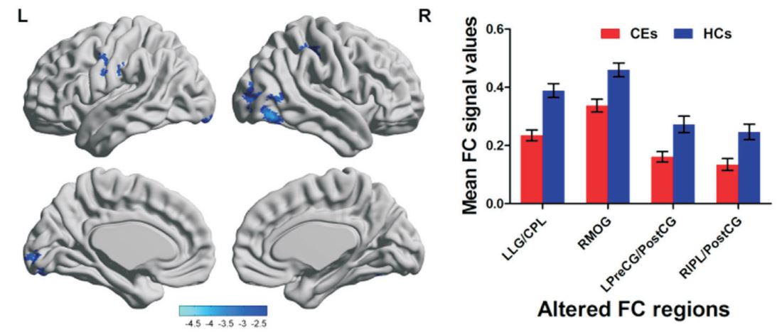
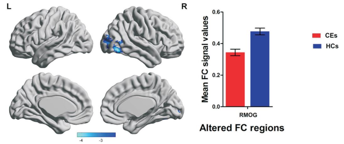
The CE patients showed significantly less FC between the left V1 and left lingual gyrus/cerebellum posterior lobe, right MOG, left precentral gyrus/postcentral gyrus and right IPL/postcentral gyrus. (Fig.1) Meanwhile, CE patients showed significantly less FC between right V1 and right MOG (BA19, 37). (Fig.2)Discussions and Conclusions
1. Our findings show that CE involves defects in the possessing of stereovision, such as fusion (lingual gyrus), spatial processing (MOG), and visual recognition (IPL)3. 2. Interestingly, significant FC decrease occurs between left V1 and frontal eye fields, suggesting the involvement of oculormotor disorder in CE4.Acknowledgements
No acknowledgement found.References
1. Chen, X., et al., Prevalence of amblyopia and strabismus in Eastern China: results from screening of preschool children aged 36-72 months. Br J Ophthalmol, 2016. 100(4): p. 515-9.
2. Yan, X., et al., Dorsal visual pathway changes in patients with comitant extropia. PLoS One, 2010. 5(6): p. e10931.
3. Li, Q., et al., Assessment of Cortical Dysfunction in Patients with Intermittent Exotropia: An fMRI Study. PLoS One, 2016. 11(8): p. e0160806.
4. Cocchi, L., et al., A hierarchy of timescales explains distinct effects of local inhibition of primary visual cortex and frontal eye fields. Elife, 2016. 5.
Figures