3273
Altered Connectivity Using Resting State fMRI in SCA1 and SCA2 PatientsDibashree Tamuli1, Shefali Chaudhary2, S. Senthil Kumaran2, Ashok Kumar Jaryal1, Achal Kumar Srivastava3, and Kishore Kumar Deepak1
1Physiology, All India Institute of Medical Sciences, New Delhi, New Delhi, India, 2NMR, All India Institute of Medical Sciences, New Delhi, New Delhi, India, 3Neurology, All India Institute of Medical Sciences, New Delhi, New Delhi, India
Synopsis
Neurodegeneration in cortical and subcortical brain areas are found in SCA type 1 and 2 patients. The structural brain volumetric analysis has been done to know the differential loss of volume of brain areas in SCA type 1 and 2. Especially, to find the functional connectivity of the whole brain in the same SCA patients, we have assessed default mode network using resting state fMRI.
Introduction
Spinocerebellar ataxia (SCA) type 1 and 2 are the autosomal dominant neurodegenerative disorders. These are the common subtypes of SCA worldwide and caused by unstable CAG trinucleotide repeat expansion at ATXN1 and ATXN2 genes for SCA type1 and 2 respectively. Cortical and subcortical atrophies (especially cerebellar) are well known in these SCAs by previous neuroimaging studies1, but very little known about the differential default mode connectivity. The use of functional connectivity (FC) measurements is a powerful tool that helps in the discrimination of neurodegenerative diseases. To know the connectivity in SCA type 1 and 2, we have studied default mode network by using resting state fMRI. Additionally, we have also explored the differential loss of volume of brain areas in the same patient groups by structural MRI.Methods
The resting state fMRI (180 dynamics) and 3D T1-weighted data were acquired using a 3T scanner (Achieva, M/s Philips Medical Systems, The Netherlands) and analyzed using conn toolbox (SPM12) in genetically proven SCA1 (n =9 , age = 35.9 ± 7.6 years) and SCA2 (n =9 , age = 30.9 ± 7.6 years) patients. ROI to ROI based analysis (p<0.05, FDR corrected) was used. While the 3D T1-weighted scans of the whole brain was analyzed by FreeSurfer (version 5.3) software in the same patient groups. MRI parameters used in T1-weighted scans were: Voxel size = 0.6×0.6×1, FOV = 240×240×180 and flip angle = 8ᵒ.Results
We found the subcortical loss of volume in medulla, pons, brainstem, left cerebellar cortex and right cerebellar cortex in SCA2 than SCA1 patients (table 1). Interestingly, a positive functional connectivity was observed in anterior cerebellum and precuneus (T =4.340; p = 0.042) and post cingulate cortex (T = 4.390; p = 0.042) in SCA1 patients in comparison to SCA2 patients.Conclusion
On the structural volumetric analysis, the cerebellar and brainstem neurodegeneration have been found to be more in SCA2 as compared with SCA1 patients. Also, the default mode network analysis may differentiate SCA type1 and 2 using fMRI. The negative connectivity in SCA2 may be attributed to the impairment in saccadic subsystem efficient for vision. The role of precuneus in spatial vision2 and posterior cingulate cortex in cognition3,4 help to understand an alternate connectivity in these patients.Acknowledgements
No acknowledgement found.References
1. Tamuli D, Kaur M, Sethi TP, Singh A, Faruq M, Jaryal AK, Srivastava AK, Kumaran SS, Deepak KK. Differential involvement of brain areas correlate with genotype and phenotype of SCA1, SCA2 and SCA3 [Communicated in Cerebellum]. 2. Cavanna AE, Trimble MR. The precuneus: a review of its functional anatomy and behavioural correlates. Brain. 2006 Jan 6;129(3):564-83. 3. Maddock RJ, Garrett AS, Buonocore MH. Posterior cingulate cortex activation by emotional words: fMRI evidence from a valence decision task. Human brain mapping. 2003 Jan;18(1):30-41. 4. Fransson P, Marrelec G. The precuneus/posterior cingulate cortex plays a pivotal role in the default mode network: Evidence from a partial correlation network analysis. Neuroimage. 2008 Sep 1;42(3):1178-84.Figures
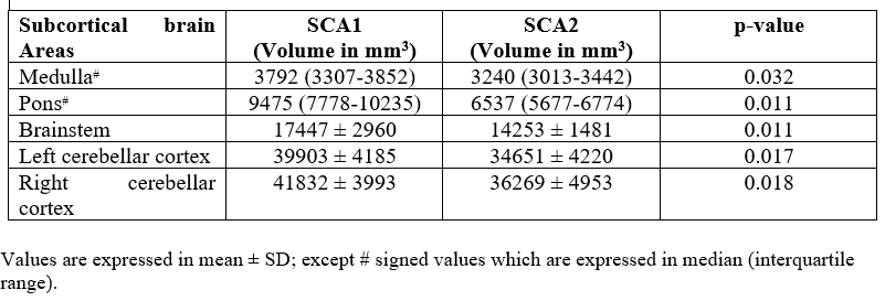
Table 1: Volume
of subcortical brain structures in
spinocerebellar ataxia type 1 and 2 patients