3272
Regularization Small-Worldization in Amyotrophic lateral sclerosis: A Resting-State fMRI Study1Department of radiology, West China Hospital of Sichuan University, Huaxi Magnetic Resonance Research Center(HMRRC), Chengdu, China
Synopsis
This study aims to use the graph-based theoretical analysis to investigate the topological properties of the functional brain connectome in amyotrophic lateral sclerosis(ALS).151 healthy controls and 101 patients underwent a rs-fMRI scan. Compared with controls, brain networks of ALS patients were characterized by decreased global efficiency and the characteristic path length increased. Based on these perspectives, the ALS group exhibited “regularization small-worldization”. A network with 10 nodes and 16 edges was identified that was significantly altered in default mode network (DMN) regions. This study may provide novel insights into the pathophysiological mechanisms underlying psychiatric disorders from a connectomic perspective.
Introduction
Amyotrophic lateral sclerosis (ALS) is a fatal, rapidly progressive neurodegenerative disease that principally affecting motor neurons[1]. Recently, both neuropathological and neuroimaging findings have provided further insight on the widespread effect of the neurodegeneration on brain connectivity in ALS[2]. However, little is known about using the graph-based theoretical analysis to investigate the topological properties of the functional brain connectome in this rare disease.Materials and methods
One hundred and fifty-five healthy controls and 101 ALS patients underwent a resting-state functional magnetic resonance imaging scan. The whole-brain functional networks were constructed based on thresholding the Pearson correlation matrices of 90 brain regions, and both global and nodal network properties were measured in graph theory approaches. Then we applied nonparametric permutation tests to identify significant between-group differences in the AUCs of all of the network metrics, to compare the small-world properties, network efficiency, and nodal characteristics of the functional connectomes between the ALS patients and the healthy controls[3]. This approach has been shown to be sensitive for detecting topological alterations of brain networks[4][5]. Then, we chose the nodes that exhibited between group differences of both nodal degree and efficiency, and created a connection matrix among those nodes for each participant and applied the network-based statistics (NBS) method to define a set of suprathreshold links among any connected components (P<0.05).Results
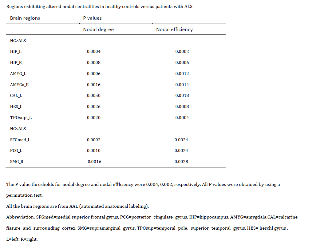
Compared with controls, brain networks of ALS patients were characterized by decreased global efficiency (Eglob) (P=0.046) and the characteristic path length (Lp) (P=0.043) increased. Based on the perspectives of segregation (Eloc) and integration (Lp),the ALS group exhibited “regularization small-worldization” in which the network transforms from a small-world network to a relatively random network[6]. Locally, compared to control subjects, patients with ALS exhibited decreased nodal degree and node efficiency in bilateral hippocampus, bilateral amygdala, calcarine fissure and surrounding cortex, heschl gyrus and superior temporal gyrus (temporal pole). Meanwhile, patients with ALS exhibited increased nodal centrality involving medial superior frontal gyrus(mSFG), posterior cingulate gyrus(PCG), supramarginal gyrus (SMG) (p<0 .05, FDR corrected). The size of the subnetwork fed into NBS was 10 × 10. A network with 10 nodes and 16 edges was identified that was significantly altered in ALS using NBS, the nodes included regions in default mode network (DMN) (PCC, mSFG, hippocampus)[7]. Significantly altered edges were observed involving each of these regions. All connectivity alterations within this network were increased in the ALS group .Discussion and Conclusions
Our analyses of topological brain networks in patients with ALS indicate the altered segregated and integrated organization[8][9]. Based on these alterd perspectives the ALS group showed a “regularization small-worldization” pattern. Furthermore, the region pairs with between-group differences of nodal characteristics showed the disequilibrium among the DMN, which might be associated with the pathophysiology of ALS. This study may provide novel insights into the pathophysiological mechanisms underlying psychiatric disorders from a connectomic perspective.Acknowledgements
This study was supported by the National NaturalScience Foundation (Grant Nos.81761128023,812 20108013, 81227002 and 81030027)References
[1] Sorrentino P, Rucco R, Jacini F, Trojsi F, Lardone A, Baselice F, Femiano C,Santangelo G, Granata C, Vettoliere A, Monsurrò MR, Tedeschi G, Sorrentino G.Brain functional networks become more connected as amyotrophic lateral sclerosis progresses: a source level magnetoencephalographic study. Neuroimage Clin. 2018Aug 4;20:564-571.
[2]Reischauer C, Gutzeit A, Neuwirth C, Fuchs A, Sartoretti-Schefer S, Weber M, Czell D. In vivo evaluation of neuronal and glial changes in amyotrophic lateral sclerosis with diffusion tensor spectroscopy. Neuroimage Clin. 2018 Oct3;20:993-1000.
[3] Bullmore, E., & Sporns, O. (2012). The economy of brain network organization.Nature Reviews, Neuroscience, 13, 336–349.
[4] Achard, S., & Bullmore, E. (2007). Efficiency and cost of economical brain functional networks. PLoS Computational Biology, 3, 174–183.
[5]Niu R, Lei D, Chen F, Chen Y, Suo X, Li L, Lui S, Huang X, Sweeney JA, Gong Q.Reduced local segregation of single-subject gray matter networks in adult PTSD. Hum Brain Mapp. 2018 Dec;39(12):4884-4892.
[6] Suo XS, Lei DL, Li LL, Li WL, Dai JD, Wang SW, He MH, Zhu HZ, Kemp GJK, Gong QG. Psychoradiological patterns of small-world properties and a systematic review of connectome studies of patients with 6 major psychiatric disorders. J Psychiatry Neurosci. 2018 Nov 1;43(6):427.
[7] Menon, V. (2011). Large-scale brain networks and psychopathology: A unifying triple network model. Trends in Cognitive Sciences, 15, 483–506.
[8]Niu R, Lei D, Chen F, Chen Y, Suo X, Li L, Lui S, Huang X, Sweeney JA, Gong Q. Disrupted grey matter network morphology in pediatric posttraumatic stress disorder. Neuroimage Clin. 2018 Mar 23;18:943-951.
[9]Suo X, Lei D, Li K, Chen F, Li F, Li L, Huang X, Lui S, Li L, Kemp GJ, Gong Q. Disrupted brain network topology in pediatric posttraumatic stress disorder: A resting-state fMRI study. Hum Brain Mapp. 2015 Sep;36(9):3677-86.
Figures