3262
Evaluate the Characteristics of Spontaneous Intracranial Artery Dissection using High Resolution MRI Vessel Wall Imaging1Department of Radiology, Changhai hospital of Shanghai, Shanghai, China, 2Changhai hospital of Shanghai, Shanghai, China
Synopsis
Compared to luminal angiographic techniques, high resolution magnetic resonance imaging is more helpful to the diagnosis and differential diagnosis the dissection from other vascular pathologies such as atherosclerosis or aneurysm. In this study, we present results obtained with vessel wall imaging to evaluate the characteristics of spontaneous intracranial artery dissection. We divided patients into two groups according to CE-MRA, and compared the characteristics of high resolution magnetic resonance imaging between two groups. The results of our study may be helpful to understand the lumen and wall change of different type of spontaneous intracranial artery dissection shown on luminal angiographic techniques.
Purpose:
High resolution MRI vessel wall imaging provides important insights of assessing both vessel lumen and wall in intracranial artery stenosis disease. This study aims to evaluate the characteristics of spontaneous intracranial artery dissection on high resolution MRI vessel wall imaging.Methods:
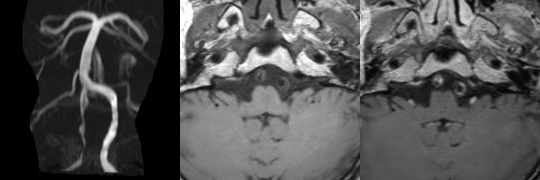
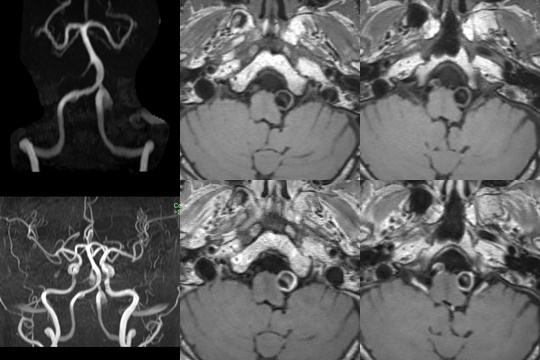
This was a retrospective study with patient consent. Thirty-four patients (7 female, age 50±14) with approved spontaneous intracranial artery dissection(DSA and clinical diagnosis) were scanned on a 3T Siemens Skyra scanner with pre- and post-contrast 3D T1-weighted SPACE (0.5mm isotropic). A high resolution CE-MRA was performed following the pre-contrast SPACE sequence. Immediately following the CE-MRA, the SPACE sequence was repeated. Patients were divided into two groups based on the artery change on CE-MRA as stenosis(occlusion) group and dilatation group. The characteristics of spontaneous intracranial artery dissection were evaluated including the location of dissection, double lumen, intimal flap, intramural hematoma(an area of hyperintense signal intensity on pre-contrast SPACE images in the vessel wall), Intraluminal contrast enhancement(an area of intraluminal contrast enhancement on post-contrast SPACE images), and artery wall enhancement(degree of enhancement was assessed based on a visual grading system as follows: grade 0, similar to that of normal vessel walls; grade 1, greater than that of grade 0 but less than or similar to that of muscle; and grade 2, greater than that of muscle). The characteristics of spontaneous intracranial artery dissection were compared between two groups. Patients clinical symptom and brain parenchyma lesion(ischemic or hemorrhage) were also evaluated and compared.Results:
Thirty patients’ dissection located on the posterior circulation. There were 16 patients in stenosis group and 18 patients in dilatation group. More patients in dilatation group showed double lumen and intimal flap. Intramural hematoma and intraluminal contrast enhancement were more frequency seen in stenosis group. There was no significantly different of artery wall enhancement between the two groups.Discussion:
Spontaneous intracranial artery dissection always show stenosis/occlusion or dilation on traditional angiography images such as computed tomography angiography (CTA), magnetic resonance angiography (MRA), and digital subtraction angiography(DSA). The definite diagnostic criteria of spontaneous intracranial artery dissection is the presence of an intimal flap and a double lumen[1]. However, in cases without a definite dissection sign, precise diagnosis may not be easy to achieve. Patients with lumenal dilatation and intramural hematoma may be misdiagnosed as intracranial aneurysm. Compared to luminal angiographic techniques, high resolution magnetic resonance imaging is more helpful to the diagnosis and differential diagnosis the dissection from other vascular pathologies such as atherosclerosis or aneurysm[2]. Intraluminal contrast enhancement and artery wall enhancement, which is suggestive of intraluminal thrombus formation, is strongly correlated with ischemic symptoms in patients with spontaneous cervical artery dissection[3]. In this study, we divided patients into two groups according to the traditional angiography images(CE-MRA), and compared the characteristics of high resolution magnetic resonance imaging between two groups. The results of our study may be helpful to understand the lumen and wall change of different type of spontaneous intracranial artery dissection shown on luminal angiographic techniques.Acknowledgements
NoneReferences
[1] Uemura M, Terajima K, Suzuki Y, et al. Visualization of the Intimal Flap in Intracranial Arterial Dissection Using High-Resolution 3T MRI. J Neuroimaging. 2017 Jan;27(1):29-32.
[2] Jung SC, Kim HS, Choi CG, et al. Quantitative Analysis Using High-Resolution 3T MRI in Acute Intracranial Artery Dissection.J Neuroimaging. 2016 Nov;26(6):612-617.
[3] Coppenrath E, Lenz O, Sommer N, et al. Clinical Significance of Intraluminal Contrast Enhancement in Patients with Spontaneous Cervical Artery Dissection: A Black-Blood MRI Study. Rofo. 2017 Jul;189(7):624-631.
Figures