3258
Reducing scan time of selective 3D TOF using the dedicated algorithm based on compressed sensing1Hitachi, Ltd., Tokyo, Japan
Synopsis
Non-contrast enhanced ICA selective 3D TOF using cylinder excitation pre-sat pulse has a possibility which visualizes blood flow from left or right ICA like DSA. It requires one set of 3D TOF scan; non-labeled 3D TOF and ICA labeled 3D TOF, then scan time becomes two times longer than conventional 3D TOF scan. In this study, compressed sensing scheme was applied to reducing the scan time of selective 3D TOF. Proposed method uses similarity between 3D TOF images with and without pre-sat pulse. This realizes only 1 minutes’ additional ICA selective 3D TOF scan to conventional 3D TOF scan.
Purpose
The purpose of this study is reducing scan time of selective 3D TOF by using compressed sensing.Introduction
ICA Selective 3D TOF1 (selective MRA) visualize blood flow from left or right ICA selectively. However, selective MRA gives additional information of blood flow of specific artery non-invasively, it has not been used routinely in clinical MRI practices. One reason is that selective MRA requires additionally another 3D TOF acquisition with pre-sat pulse to saturate blood signal from specific artery, and the scan time is doubled. In this study, compressed sensing2 was applied to selective MRA. The algorithm uses information of 3D TOF without pre-sat pulse that is acquired almost all clinical routine MRI practices. The scan time becomes shorter to about 1/5 times compared to without using compressed sensing. We demonstrated in-vivo experiment and showed its image quality is comparable to that of without compressed sensing.Materials and Methods
Experiments
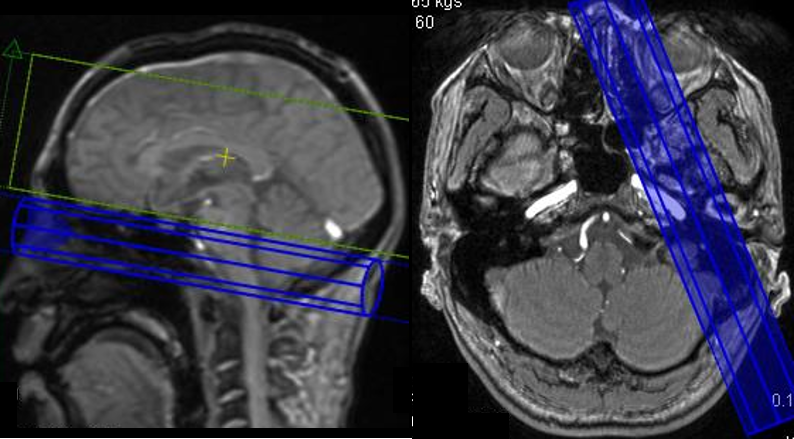
Experiment was conducted on 3 Tesla whole body MRI system (Hitachi, Ltd.). A healthy volunteer was evaluated. This study was approved by the ethics committee of Hitachi group headquarters. 15-channel receive coil was used. Scan parameters of a 3D TOF scan were as follows; TR / TE = 23.8 / 3.3 ms, FA = 15°, thickness = 1.2 mm, FOV = 220 mm, Freq# x Phase# x slice# = 320 x 224 x 60, reconstruction matrix = 512 x 512 x 120, and scan time was 5 min 20 sec. Two 3D TOF images (with pre-sat pulse, and without pre-sat pulse) were acquired. And total scan time was 10 min 40 sec. In this study, pre-sat pulse was set at left ICA and blood signal from right ICA was imaged. The position of pre-sat pulse is shown in Figure 1. Sampling and Reconstruction algorithm
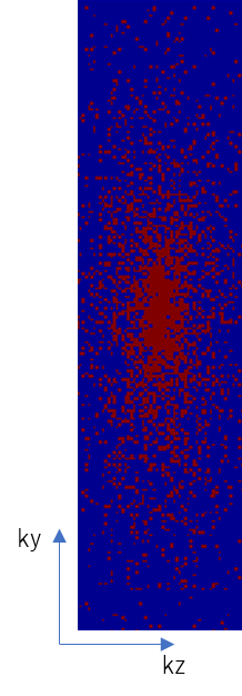
The k-space data of selective MRA was under-sampled retrospectively. Variable density random sampling was used. Under-sampling factor was set to 5.3. Sampling pattern is shown in Figure 1. The scan time corresponds to 1 min. In image reconstruction, the l1 norm in equation 1 was minimized.
$$I_{wSAT}^i= argmin (|(|F_u I_{wSAT}^i-y^i|)|_2^2+λ|I_{woSat}^i-I_{wSat}^i |_1)$$(1)
Where, is under sampled Fourier transform, is image of selective 3D TOF, y is sampled k-space data, is image of 3D TOF without pre-sat pulse, and i is index of receive coil channels (i = 1-15). The first term in equation 1 denotes data consistency, and the second term denotes the assumption that difference between two 3D TOF images those acquired with and without pre-sat pulse is sparse. ADMM (Alternating Direction Method of Multipliers) algorithm3 were used to minimize equation 1. Iteration was set 20 times and reconstruction time was 15 min.
Combine multi-channel data
The reconstructed image of each receive channel were combined as usual sum of square algorithm in equation 2.
$$I_{wSAT}=\sqrt{∑_{i=1}^{15}(I_{wSAT}^i )^2}$$ (2)
Evaluation Signal intensity of saturated blood signal, visualized blood signal, and peripheral vessel was compared between fully sampled image, and reconstructed image using proposed algorithm.
Results and Discussions
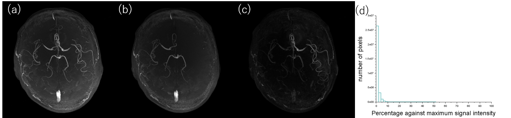
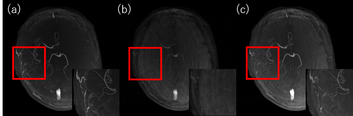
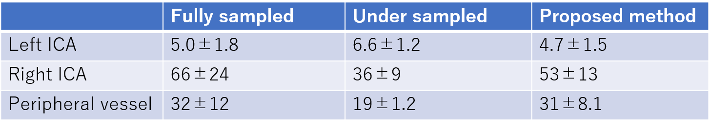
Figure 3 shows MIP image of 3D TOF image (a), selective MRA (b), and difference of these two images (c). Histogram of difference image is also shown in (d). Signal of over 70 % of pixels were less than 1 % of maximum signal difference, and the difference image was sparse. Figure 4 shows MIP image of selective MRA acquired with fully sampled (a), under-sampled (b) and reconstructed using proposed method (c). However small arteries were not observed in under-sampled image due to artifacts caused by under-sampling, by using proposed reconstruction algorithm, small arteries were reconstructed clearly. In Table 1, signal intensity of left and right ICA and peripheral vessel are shown. Signal intensity in under-sampled image of right ICA and peripheral vessel was smaller than fully sampled or proposed reconstruction, due to artifact in under-sampling image. The proposed compressed sensing algorithm that uses the information of 3D TOF without pre-sat pulse worked well because the difference image was sparse, and only 1/5 acquisition was enough to acquire selective MRA.Conclusions
In this study, compressed sensing scheme was applied to reducing the scan time of selective 3D TOF. Proposed method uses similarity between 3D TOF images with and without pre-sat pulse. This realizes selective 3D TOF images can be acquired with additional only 1 minutes’ scan. This technique will be useful for fast scanning of selective 3D TOF images.Acknowledgements
No acknowledgement found.References
T. Nishihara et al. ISMRM 2012; 20: 2497.
M. Lustig et al. Magnetic Resonance in Medicine, 58 (2007) 1182-1195.
S. Boyd et al. Foundations and Trends in Machine Learning, 3 (2010) 1.
Figures