3257
in vivo quantitative assessment of the meningeal lymphatics using 3D black blood T1 imaging: a preliminary study1Gangnam Severance Hospital, Seoul, Korea, Republic of
Synopsis
We evaluated the meningeal lymphatics using 3D black blood T1 imaging and its association with clinical parameters as well as enlarged perivascular spaces. This retrospective study included 24 patients who underwent contrast-enhanced 3D black blood T1 imaging on 3T brain MRI and assessed their meningeal lymphatics located parallel to the superior sagittal sinuses. The group with higher meningeal lymphatics volume more frequently have diabetes than the lower group, which is one of the major vascular as well as cognitive risk factors. Furthermore, the higher group had a significantly higher score of enlarged perivascular space in centrum semovale, which is not surprising when considering its close anatomical and functional relationships within the glymphatic system. Therefore, we suggest that the expanded meningeal lymphatics may be an imaging marker of poor function of glymphatics but further studies with disease conditions such as Alzheimer’s disease should be followed to clarify the exact meaning of the volume of the meningeal lymphatics.
Synopsis
We evaluated the meningeal lymphatics using 3D black blood T1 imaging and its association with clinical parameters as well as enlarged perivascular spaces. This retrospective study included 24 patients who underwent contrast-enhanced 3D black blood T1 imaging on 3T brain MRI and assessed their meningeal lymphatics located parallel to the superior sagittal sinuses. The group with higher meningeal lymphatics volume more frequently have diabetes than the lower group, which is one of the major vascular as well as cognitive risk factors. Furthermore, the higher group had a significantly higher score of enlarged perivascular space in centrum semovale, which is not surprising when considering its close anatomical and functional relationships within the glymphatic system. Therefore, we suggest that the expanded meningeal lymphatics may be an imaging marker of poor function of glymphatics but further studies with disease conditions such as Alzheimer’s disease should be followed to clarify the exact meaning of the volume of the meningeal lymphatics.Purpose
Recently, the meningeal lymphatics have been introduced as a network of lymphatic vessels located parallel to the dural sinuses and meningeal arteries.1,2 As a part of the glymphatic system, the meningeal lymphatics are responsible for draining immune cells, small molecules, and excess fluid from the CNS.1,2 As a previous study identified the meningeal lymphatics using 3D black blood T1 imaging,3 we performed a quantitative approach to the meningeal lymphatics on 3D black blood T1 imaging and its relationship with perivascular spaces, which may offer a better understanding of the glymphatic system.Methods
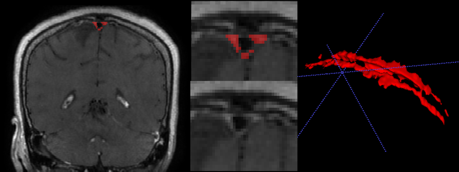
We analyzed 24 patients who underwent brain MRI from April to May 2018 for the evaluation of brain metastasis but had no visible intracranial lesion on MRI. 3D black blood T1 imaging was implemented using a fast spin echo sequence, with TR = 500 ms, TE = 24.5 ms, flip-angle = 90° and BW = 488 Hz/pixel. Images were acquired with a 256×224 matrix, 22×22 cm2 FOV, and 1.0mm thickness after injection of a gadolinium contrast agent (0.2 mmol/kg gadobutrol), using the 3T MR scanner (GE 750) with a 16-channel phased array coil. The meningeal lymphatics were identified on coronal reconstructed images as gadolinium enhancing area located parallel to the superior sagittal sinuses. A neuroradiologist manually segmented the meningeal lymphatics by using the ITK-SNAP software. Cerebral perivascular spaces in basal ganglia, centrum semiovale, and midbrain were qualitatively rated on axial T2-weighted images by using the followed scoring system: Basal ganglia and centrum semiovale perivascular spaces were rated 0 (none), 1 (1-10), 2 (11-20), 3 (21-40) and 4 (>40), and midbrain perivascular spaces were rated 0 (none visible) or 1 (visible).4 Fisher’s exact test was applied to compare the frequency of categorical variables and the Mann–Whitney U test was used to compare continuous variables.Results
There were 13 men and 11 females with a median age of 61 years (IQR, 56 – 73). The median volume of superior sagittal meningeal lymphatics was 3039 ml (IQR, 2034 – 3475.3). The patients were divided into lower meningeal lymphatic volume group (n = 13, M:F = 8:5, median age = 60 years (IQR, 59.5 – 70.5)) and higher meningeal lymphatic volume group (n = 11, M:F = 8:3, median age = 66 years (IQR, 58 – 74)) by the median value of superior sagittal meningeal lymphatics. The higher group were more likely to have diabetes mellitus (P = 0.031) than lower group while the incidence of other clinical factors of hypertension, dyslipidemia, past history of cerebrovascular disease or heart disease was not statistically significant. Qualitative perivascular space score of centrum semiovale was higher in higher meningeal lymphatic volume group than in a lower group (3 (IQR, 2 – 4) vs. 2 (IQR, 1 – 3), P = 0.030).Acknowledgements
N/AReferences
1. Jessen NA, Munk AS, Lundgaard I, Nedergaard M. The Glymphatic System: A Beginner's Guide. Neurochem Res. 2015.40(12):2583-2599.
2. Louveau A, Smirnov I, Keyes TJ, et al. Structural and functional features of central nervous system lymphatic vessels. Nature. 2015.523(7560):337-341.
3. Absinta M, Ha SK, Nair G, et al. Human and nonhuman primate meninges harbor lymphatic vessels that can be visualized noninvasively by MRI. Elife. 2017.6.
4. Potter GM, Chappell FM, Morris Z, Wardlaw JM. Cerebral perivascular spaces visible on magnetic resonance imaging: development of a qualitative rating scale and its observer reliability. Cerebrovasc Dis. 2015.39(3-4):224-231.
Figures