3250
Pulsed ASL prepared 4-Dimensional Dynamic Intracranial MR Angiography at 7T with Improved B1 and B0 robustness1Siemens Medical Solutions USA, Inc., Los Angeles, CA, United States, 2Keck school of medicine, University of Southern California, Los Angeles, CA, United States
Synopsis
This work enhances the quality of non-contrast enhanced time-resolved 4-dimensional MR angiography (4D-MRA) at 7T, by combining segmented multi-phase acquisition of the FAIR scheme with labeling pulses of improved robustness to B1/B0 inhomogeneity and chemical shift. The gradient-modulated offset independent inversion pulses were optimized for 7T application maximizing MR system performance, while providing uniform inversion, sharp transition and tolerance to chemical shift. 4D-MRA images with higher spatial-temporal resolution were reliably acquired within SAR limit of normal operation mode within 5 minutes, presenting a valuable tool for assessing intracranial vasculature and hemodynamics.
Purpose
Non-contrast enhanced time-resolved 4-dimensional MR angiography (4D-MRA) is valuable for assessing intracranial vasculature and hemodynamics[1], [2]. Ultra-high-field (UHF) 7T benefits this technique with prolonged blood T1 relaxation and inherently higher SNR. However, the B0/B1 inhomogeneity often compromises the performance of inversion pulse, giving rise to imperfect inversion profile, reduced labeling efficiency and residual background signal. In this study, we optimized gradient-modulated offset independent (GOIA) inversion pulse [3] at 7T for improved B0/B1 performance and compared it with conventional hyperbolic-secant (HS) [4] and wideband-uniform rate-smooth truncation (WURST) [5] inversion pulses in 4D-MRA.Method
The FAIR labeling scheme was adopted here, which offers higher SNR than STAR scheme as previously reported. The conventional adiabatic full passage (AFP) pulses typically used in FAIR include HS and its variant HSn, and WURST. HSn and WURST are popular choices providing flat profiles to avoid RF amplitude clipping (common with HS at UHF). These AFPs typically have bandwidths of several thousand Hz, limited by the pulse duration, T2-relaxation, available peak RF amplitude, and specific absorption rate (SAR). A desired inversion thickness is achieved by adjusting the amplitude of the constant slice-selection gradient. Instead of applying constant slice-selection gradient, the GOIA pulse enhances its performance by modulating gradient waveforms. The GOIA pulse is similar to FOCI pulse in principle [6], but provides offset-independent adiabaticity (OIA), capable of uniformly rotating longitudinal magnetization within the desired bandwidth.
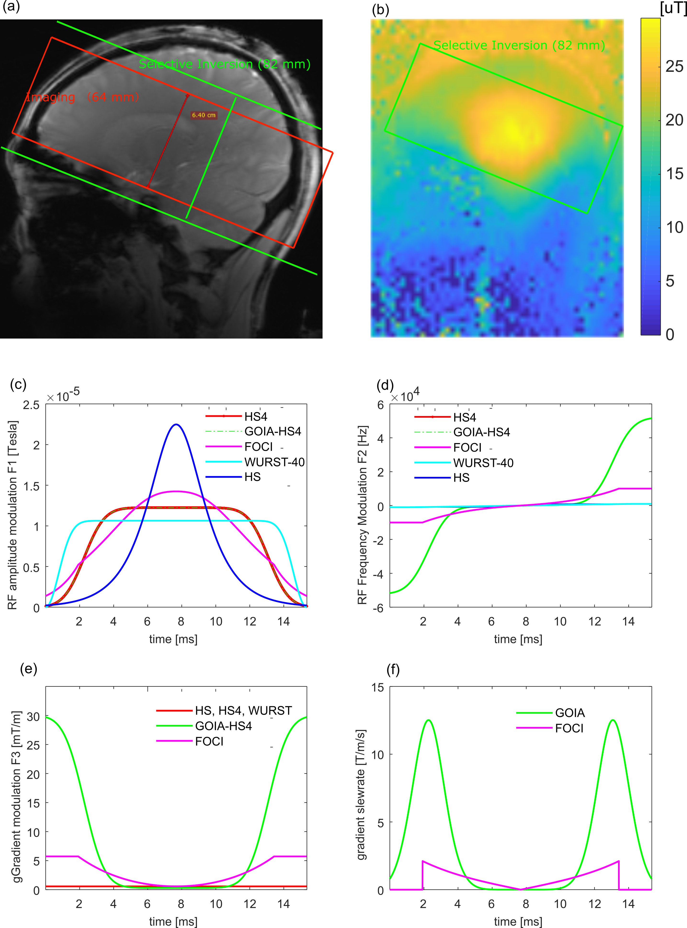
A discrete time event-based Bloch simulator [7] was implemented to numerically evaluate the performance of these pulses. The modulation functions and inversion profiles of several AFP pulses are shown in Fig.1. Here, FOCI was constructed by scaling HS using a common time-variant profile (magenta, Fig.1e). The GOIA pulse was designed based on HS4 RF amplitude modulation ($$$F_{1}$$$), and an analytical gradient modulation $$F_{3}=G_{max}[1-\rho sech\left ( \beta \tau ^{2} \right )]$$, where $$$\rho$$$ is normalized time variable, $$$G_{max}$$$= maximum gradient strength, and $$$sech\left ( \beta \tau ^{2} \right )=0.01$$$. The frequency modulation ($$$F_{2}$$$) was derived satisfying the differential equation [3], $$\frac{dF_{2}}{d\tau }-\frac{F_{2}}{F_{3}}\frac{dF_{3}}{d\tau }=\frac{F_{1}^{2}}{\kappa }$$ Multiple sets of $$$\rho$$$ and $$$\kappa$$$ were designed according to the desired slice thickness, while maximizing $$$G_{max}$$$.
Multi-phase segmented spoiled-GRE sequence was used in 4D-MRA with different types of inversion pulses. 5 healthy volunteers were recruited for imaging on a 7T system (MAGNETOM Terra, Siemens Healthineers) using 1-transmit/32-receive head coil (Nova Medical, MA, USA). The imaging parameters include: voxel size=1×1×1mm3, 64slices, FA=9°, 9 phases with temporal resolution of 100ms, scan time=4mins. Additionally, super-high resolution 4D MRA images (0.5×0.5×0.5mm3) using GOIA was also collected on two of the five subjects.
Results and Discussion
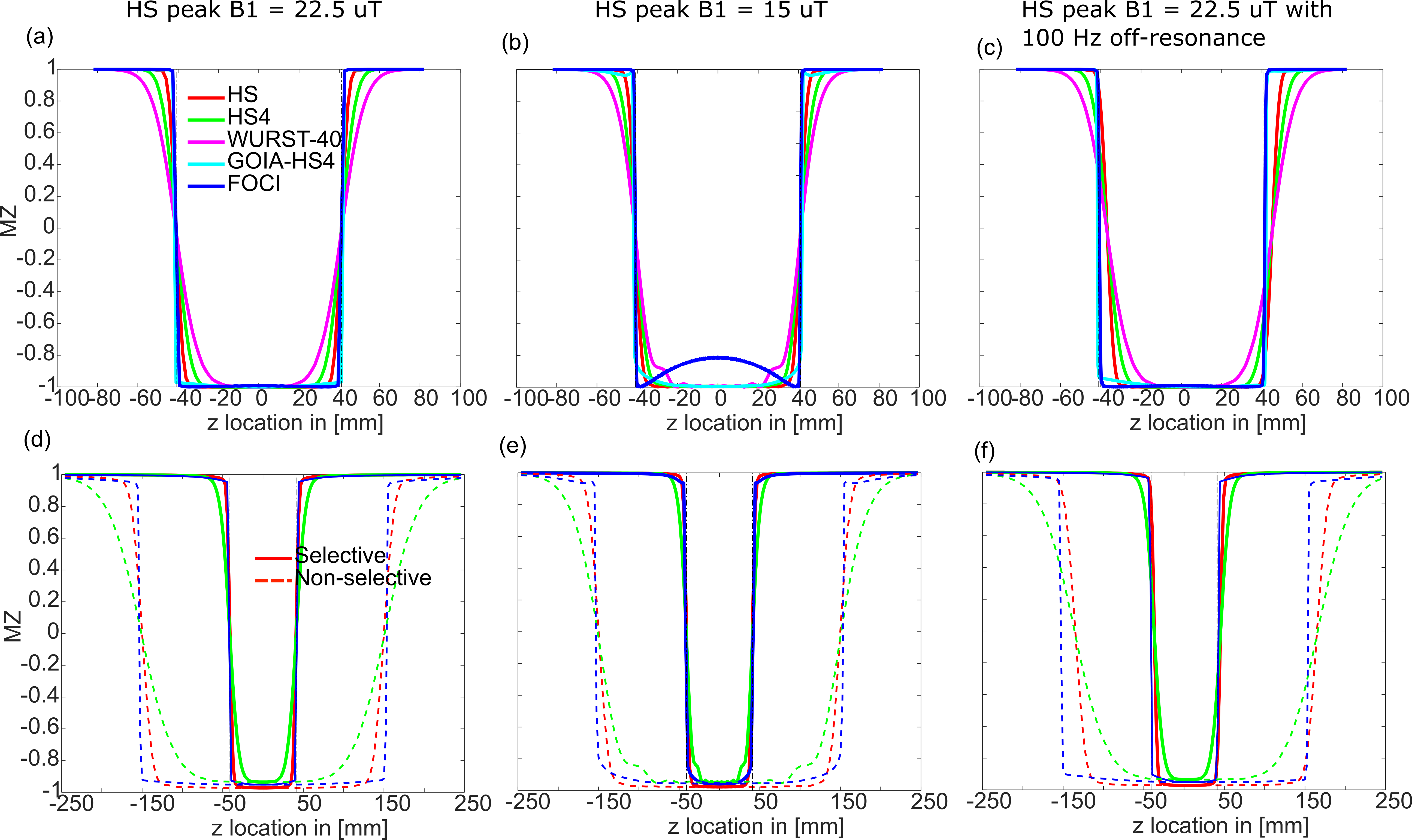
Figs.2a-c show the simulated inversion profiles. With adequate B1 amplitude (Fig.2a), all pulses achieved complete inversion, whilst GOIA and FOCI provided extremely sharp transition. WURST-40 used lowest peak RF amplitude, however its transition was the widest. With lower peak B1 at 15 uT (Fig.2b), FOCI and WURST-40 pulses’ inversion profiles deteriorated noticeably. GOIA and FOCI had superior resistance to off-resonance (Fig.2c), while conventional AFP pulses presented slice shift proportional to off-resonance. Overall, GOIA pulse had superior robustness to B0/B1 inhomogeneity compared to all the other pulses. Due to non-uniform inversion of FOCI, and similar performance between HS4 and WURST-40, FOCI and HS4 were excluded from imaging experiments.
When considering T2-relaxation (Figs.2d-2f), GOIA pulse had slightly less inversion efficiency compared with HS. However, GOIA provides significantly larger bolus as represented by the size of non-selective inversion and significantly better off-resonance performance.
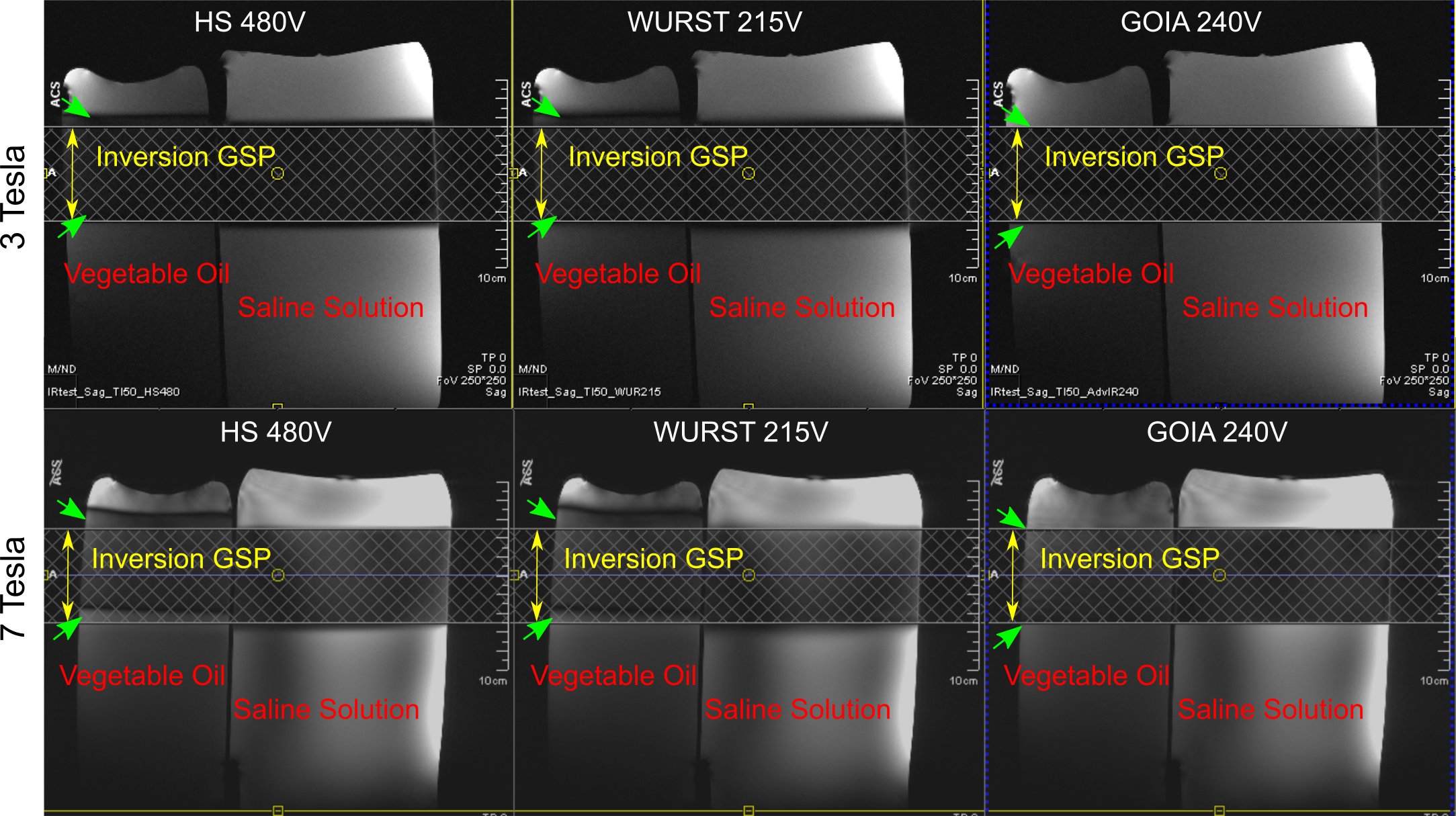
Fig.3 verifies the predicted advantages of GOIA pulse (e.g., sharp transition, uniform inversion and robustness to chemical shift) in fat-water phantom experiments at 3T and 7T. More pronounced B1/B0 inhomogeneity and more significant chemical shift indicated the necessity of better inversion pulses at 7T.
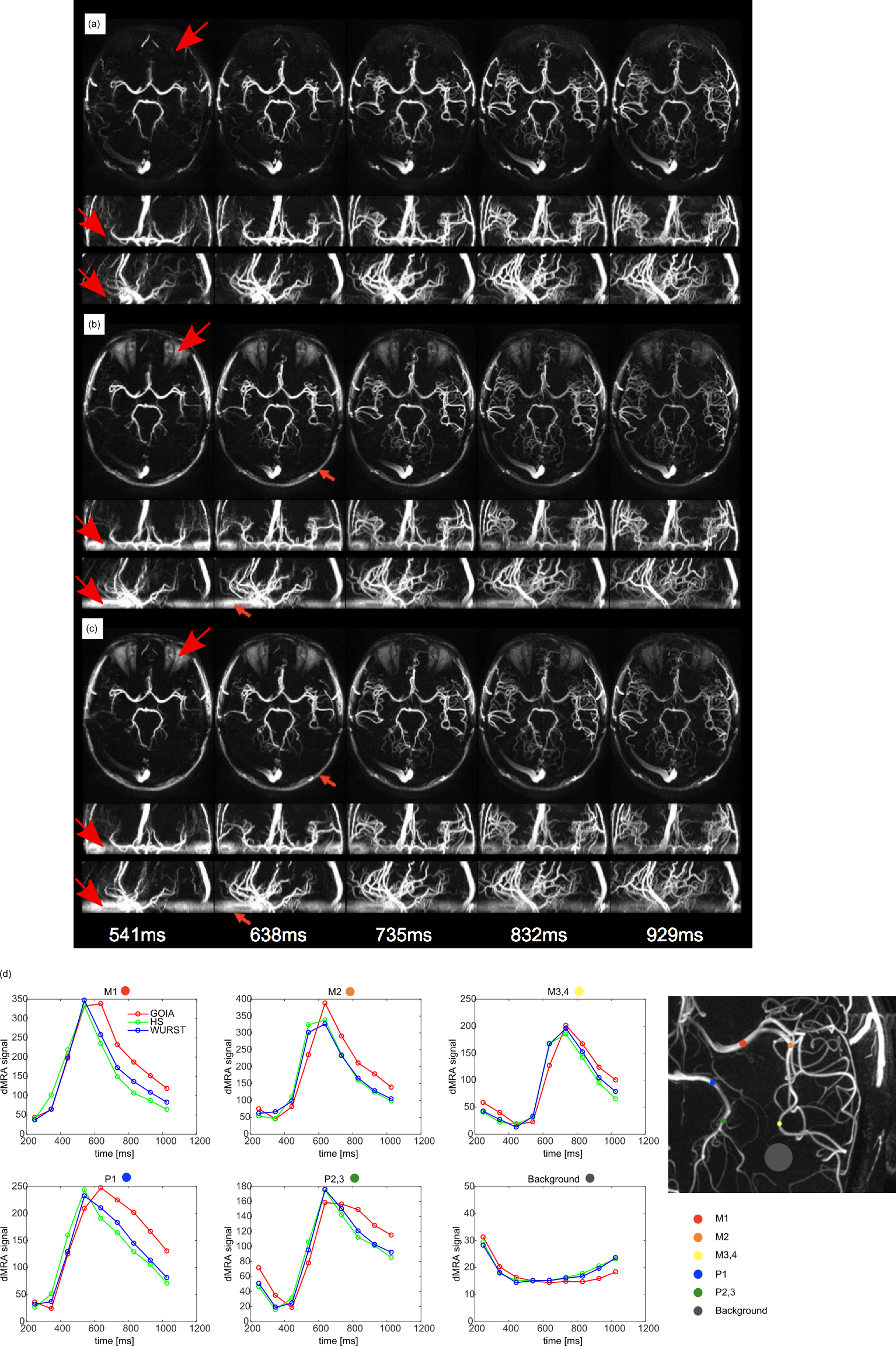
Fig.4 shows the representative 4D-MRA MIP images using different inversion pulses with a common duration of 15 ms and equal SAR. Fat-water shift was clearly visible with HS and WURST-40 as shown by the arrows, but absent in GOIA images. Better depiction of vessels with clearer background was obtained with GOIA along all three orthogonal directions. No saturation pulse was used because of SAR restriction (normal operation mode). However, background was free of residual signals. MRA signals were extracted from different vascular segments and we can see stronger MRA signal especially at latter phases with GOIA, as predicted by the simulations.
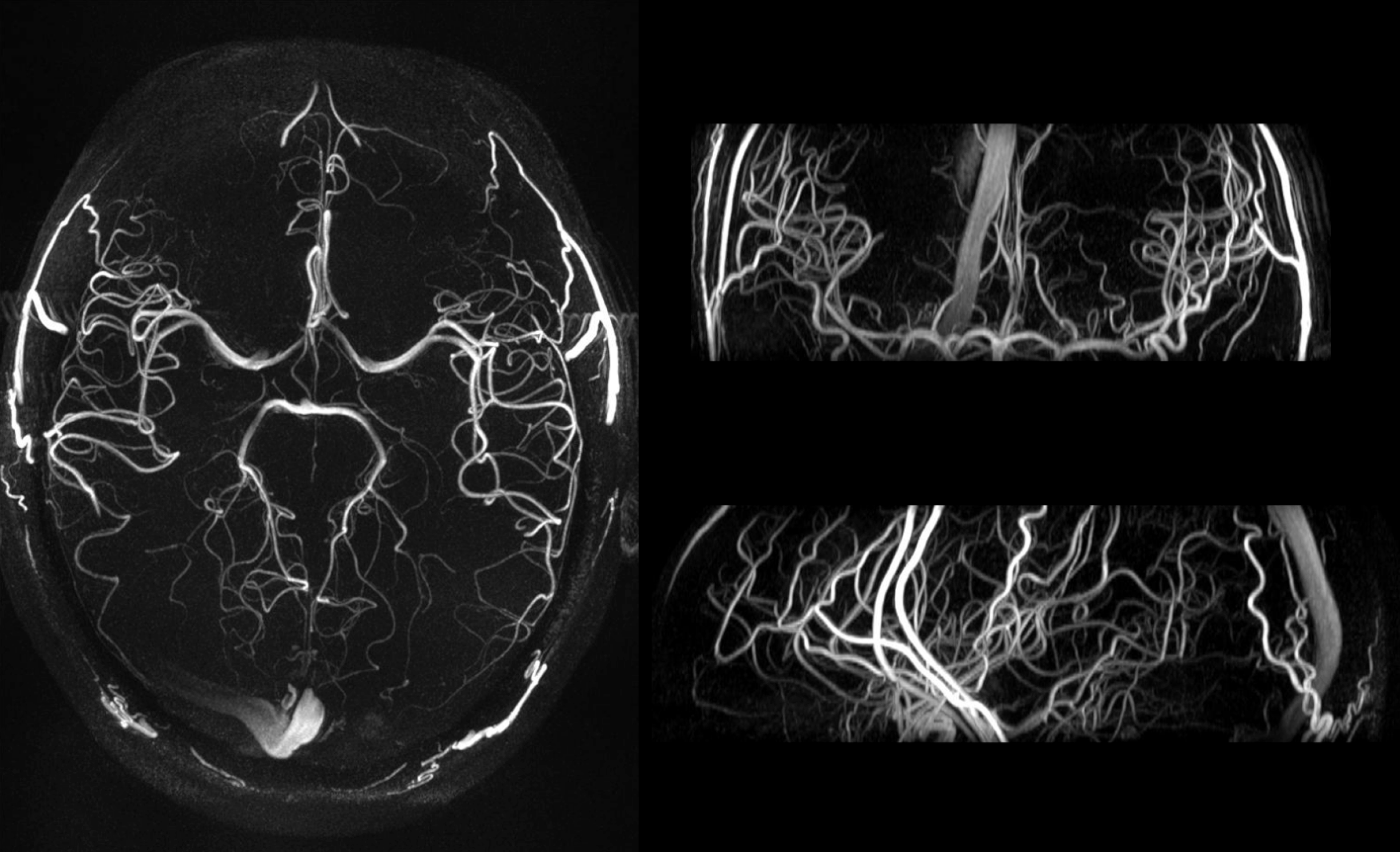
Fig.5 shows collapsed MIP images at significantly higher spatial resolution of 0.5x0.5x0.5 mm3 and reduced temporal resolution at 140ms. One can appreciate very good delineation of vasculature at all vessel sizes, which further demonstrated the capability and potential of 4D-MRA at UHF.
Conclusion
This study demonstrated that optimized gradient-modulated offset independent adiabatic (GOIA) pulses can improve image quality of 4D-MRA at 7T with improved robustness to B0/B1 inhomogeneity.Acknowledgements
AHA16SDG29630013
NIH K25AG056594
References
[1] X. Bi, P. Weale, P. Schmitt, S. Zuehlsdorff, and R. Jerecic, “Non-contrast-enhanced four-dimensional (4D) intracranial MR angiography: A feasibility study,” Magn. Reson. Med., vol. 63, no. 3, pp. 835–841, 2010.
[2] L. Yan and J. Wang, “Angiography : High Spatial and Temporal Resolution by Using True FISP – based Spin Tagging with Alternating Radiofrequency,” Radiology, vol. 256, no. 1, 2010.
[3] A. Tannús and M. Garwood, “Adiabatic pulses,” NMR Biomed., vol. 10, no. 8, pp. 423–434, 1997.
[4] M. S. Silver, R. I. JOSEPH, and D. I. HOULT, “Highly Selective Pi/2 and Pi Pulse Generation,” J. Magn. Reson., vol. 59, pp. 347–351, 1984.
[5] Ē. Kupče and R. Freeman, “Adiabatic Pulses for Wideband Inversion and Broadband Decoupling,” Journal of Magnetic Resonance, Series A, vol. 115, no. 2. pp. 273–276, 1995.
[6] R. J. Ordidge, M. Wylezinska, J. W. Hugg, E. Butterworth, and F. Franconi, “Frequency offset corrected inversion (FOCI) pulses for use in localized spectroscopy,” Magn. Reson. Med., vol. 36, no. 4, pp. 562–566, 1996.
[7] R. K. S. Kwan, A. C. Evans, and B. Pike, “MRI simulation-based evaluation of image-processing and classification methods,” IEEE Trans. Med. Imaging, vol. 18, no. 11, pp. 1085–1097, 1999.
Figures