3248
Non-contrast 3D Black Blood MRI for Intracranial Aneurysm Surveillance: A Quantitative Study1University of California San Francisco, San Francisco, CA, United States
Synopsis
Patients with unruptured intracranial aneurysms routinely undergo surveillance imaging to monitor their growth.
Purpose
CE-MRA shows good accuracy in intracranial aneurysm size measurement. However, it is undesirable for repeated imaging because of the use of contrast agents. 3D non-contrast black blood MRI has high image resolution with good visualization of aneurysm geometry and also provides evaluation of the aneurysm wall1. However, it has not been validated quantitatively against CE-MRA for aneurysm volume measurement. This study aims to compare black blood MRI with CE-MRA for aneurysm volume measurements.Methods
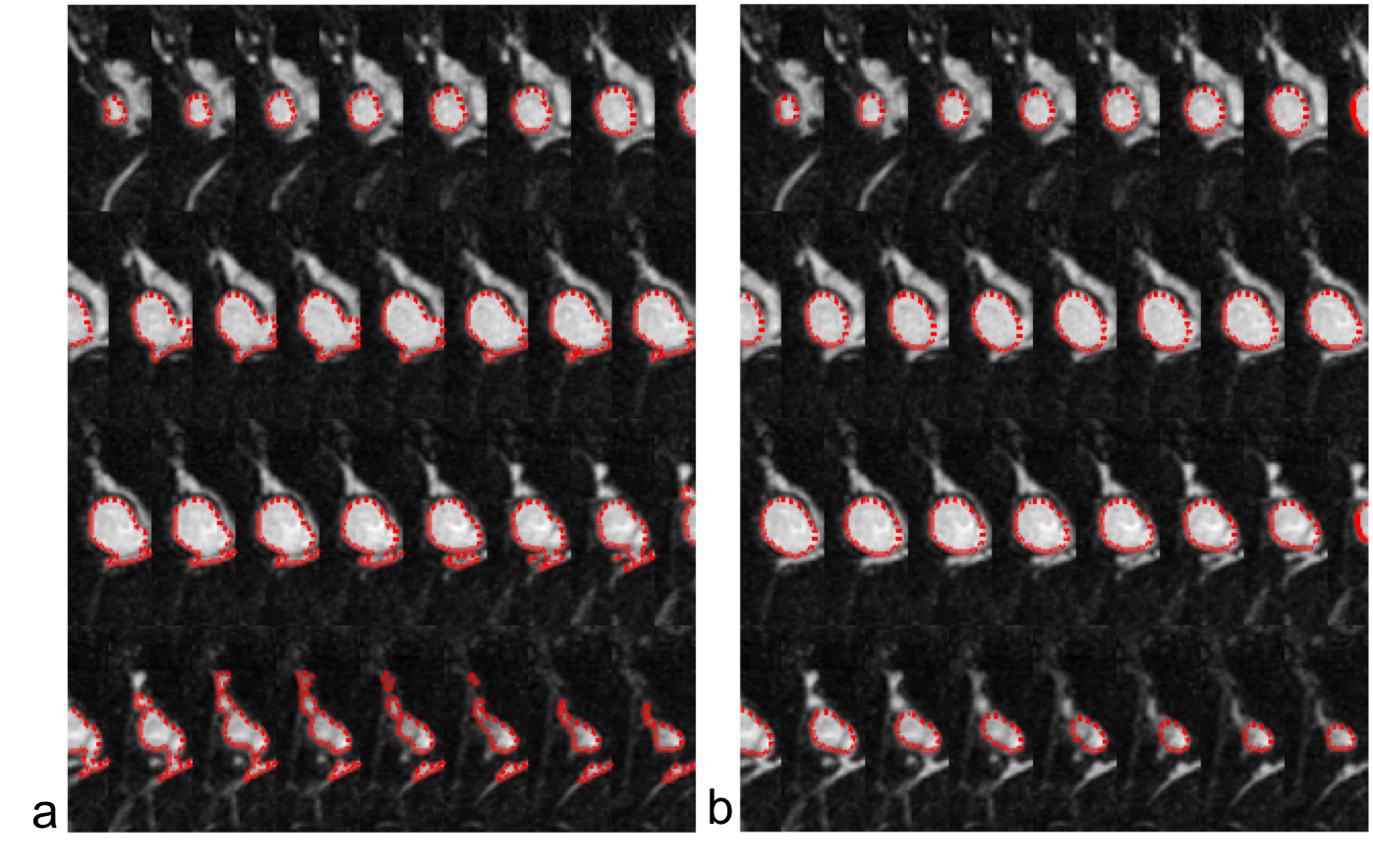
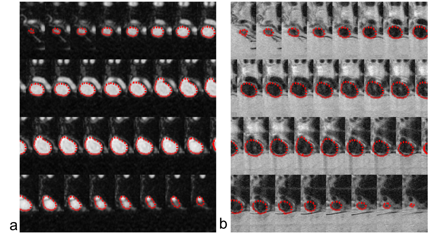
(a) Study population: 17 aneurysms from 17 patients were included in this study. All patients were scanned in a Siemens 3T scanner (Skyra) using the standard head coil. (b) Sequences: 1) clinical CE-MRA was acquired in the coronal plane using first pass with Gd-DTPA, isotropic resolution of 0.7mm, scan time 30 seconds; 2) 3D black blood SPACE (variable flip angle fast-spin-echo was acquired in the sagittal plane, 0.5mm isotropic resolution, echo train length 60, TR/TE = 900/5.6ms, scan time ~10min. (c) Image segmentation: fully automatic segmentation of intracranial aneurysms is difficult because the aneurysms, especially small aneurysms, are hard to localize. We propose a segmentation algorithm that uses a given initial contour for the first image, and then automatically segments the aneurysm using an elliptically refined level set method for the remaining images. The elliptically refined level set adds a shape prior term in the classic level set equation to avoid the contour leakage phenomena. The energy formulation of this model is:
$$E=E_{level set}+\alpha E_{ellipse}$$
where $$$E_{level set}$$$ is the data attachment term and $$$E_{ellipse}$$$ embeds the shape prior. For the data attachment term, we used the chassis Chan-Vese model2:
$$E_{level set}(c_1,c_2,C)=\mu Length(C) + \nu Area(inside(C)) +\omega_{1} \int_{inside(C)}|I-c_1|^2dxdy+\omega_{2} \int_{outside(C)}|I-c_2|^2dxdy$$
where $$$\mu$$$, $$$\nu$$$, $$$\omega_1$$$ and $$$\omega_2$$$ are fixed positive parameters. I is the intensity of the image, and $$$c_1$$$ and $$$c_2$$$ are the average intensity inside and outside $$$C$$$, respectively.
Results and discussion
The results obtained from 17 aneurysms found that the segmentation from black blood MRI was comparable with CE-MRA. The volumetric difference is 3.00%±1.72%. The proposed method can be successfully used to avoid the 3%~10% inter- and intra- observer variance that occurs in manual segmentation. Our results support the use of non-contrast black blood MRI to replace CE-MRA for the monitoring of intracranial aneurysms. Considering the recent concerns of Gadolinium brain and bone deposition, non-contrast techniques have high potential as a tool for aneurysm surveillance. The SPACE sequence also provides evaluation of the aneurysm wall, which is a unique advantage. Vessel wall features have been studied as potential markers for aneurysm rupture3.Conclusion
The proposed method is reproducible and reliable for intracranial aneurysm segmentation from 3D black blood MRI and CE-MRA. Based on our semi-automatic segmentation, the 3D black blood MRI achieves comparable accuracy to CE-MRA for intracranial aneurysm volume measurement.Acknowledgements
This study is supported by NIH grants R01HL114118 (DS), R01NS059944 (DS), and K25EB014914 (JL).References
1. Zhu CC, Wang XR, Tian B, et al. Surveillance of unruptured intracranial saccular aneurysms using non-contrast 3D black blood MRI: comparison of 3D TOF and CE-MRA with DSA.” ISMRM, Paris, 2018. 2. Chan, Tony, and Luminita Vese. "An active contour model without edges." International Conference on Scale-Space Theories in Computer Vision. Springer, Berlin, Heidelberg, 1999. 3. Edjlali M, Gentric JC, Regent-Rodriguez C, Trystram D, Hassen WB, Lion S, et al. Does aneurysmal wall enhancement on vessel wall mri help to distinguish stable from unstable intracranial aneurysms? Stroke. 2014;45:3704-3706Figures