3242
Highly Accelerated Whole Brain Isotropic 0.5mm Intracranial Vessel Wall Imaging with Nonlocal Denoising and Super Resolution Enhancement1Philips Research North America, Cambridge, MA, United States, 2Department of Radiology, University of Washington, Seattle, WA, United States, 3Department of Surgery, Division of Vascular Surgery, University of Washington, Seattle, WA, United States, 4Philips Research Hamburg, Hamburg, Germany, 5Center for Biomedical Imaging Research, Department of Biomedical Engineering, Tsinghua University, Beijing, China
Synopsis
The three-dimensional (3D) turbo spin echo (TSE) sequence with variable flip angle (VFA) has proven to be clinically useful for high resolution intracranial vessel wall imaging (VWI) at 3T. In this study, a T1 weighted 3D TSE sequence was optimized for whole brain isotropic 0.5mm intracranial VWI in 8mins28secs. The optimized VFA design improves the flow suppression in small vessels and the nonlocal denoising effectively reduces the noise amplification after super resolution enhancement while improving the vessel wall boundary definition. This combined imaging and post-processing technique provides a promising tool for intracranial atherosclerosis and stroke investigation.
Introduction
The three-dimensional (3D) variable flip angle (VFA) turbo spin echo (TSE) sequence has been commonly used for high resolution intracranial vessel wall imaging (VWI)1-5. A recent signal-to-noise ratio (SNR) priority VFA design strategy combined with the super resolution approach6 has demonstrated great potential for clinical use by improving SNR, image sharpness and scan efficiency. A remaining hurdle for routine patient scans has been the limited scan coverage which is usually a thin slab that cannot cover all major intracranial arteries. In addition, the super resolution method designed to improve the sharpness of vessel wall and plaque boundaries may enhance the background noise as a side effect. In this study, the T1 weighted 3D TSE sequence was further optimized to achieve whole brain isotropic 0.5mm intracranial VWI in 8mins28secs, and a nonlocal neural network (NN)7 based image denoising module was integrated with the super resolution post-processing to reduce the background noise amplification and further improve the overall image quality.Methods
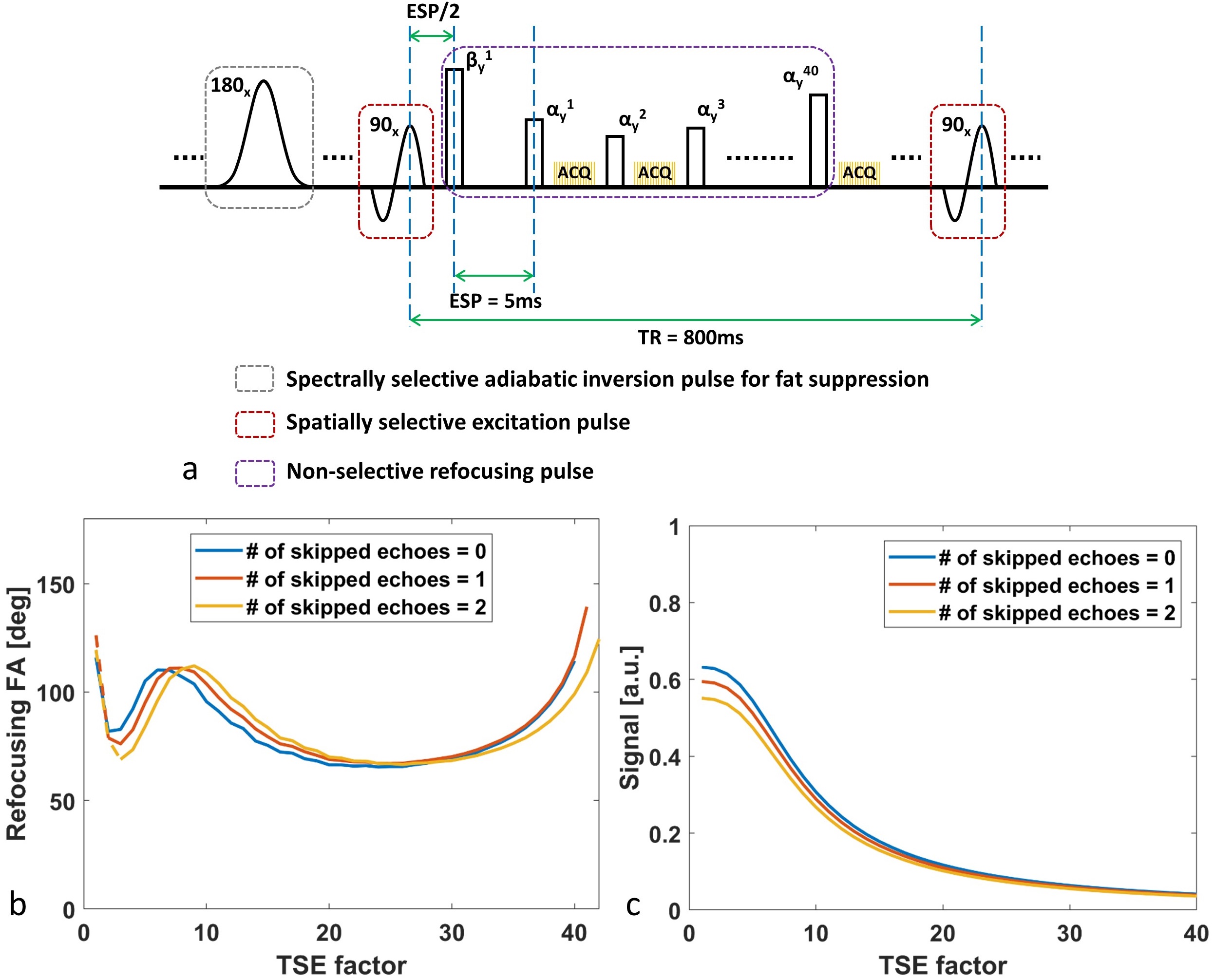
T1 weighted 3D TSE sequence optimization: A similar VFA design strategy6 was exploited to achieve a low-pass filtering shaped point spread function during TSE signal evolutions and the echo train length of 40 was chosen as a compromise between SNR gain and scan efficiency. In addition, one or two first echoes were skipped to compare with a non-skipping echo scheme in terms of the flow suppression performance in small arterial branches. The corresponding first several refocusing pulses during the initial echo skipping transition period were designed to minimize the impact on the SNR penalty in comparison to its counterpart without echo skipping (figure 1).
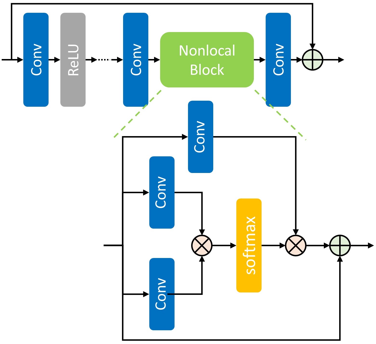
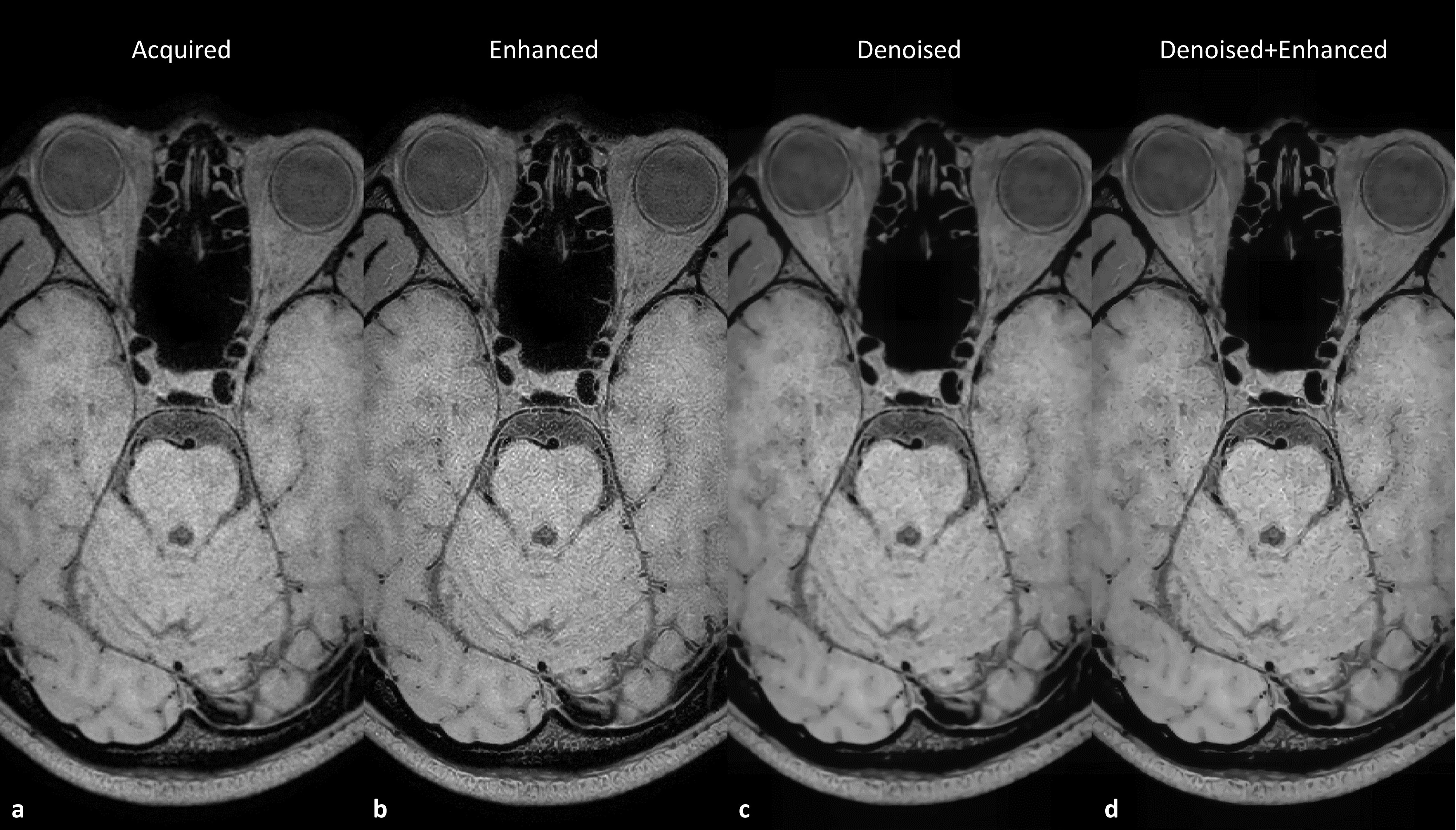
Image enhancement with noise reduction and super resolution: Inspired by the nonlocal mean method8, a residual learning based nonlocal NN7 was trained for image denoising using the nonlocal self-similarity property (figure 2). After nonlocal image denoising to suppress the incoherent background noise, the image was processed using another residual learning based convolutional NN for super resolution processing6 to improve the image sharpness. The training and testing of NN models were implemented with tensorflow on a single GPU (Nvidia, Titan Xp).
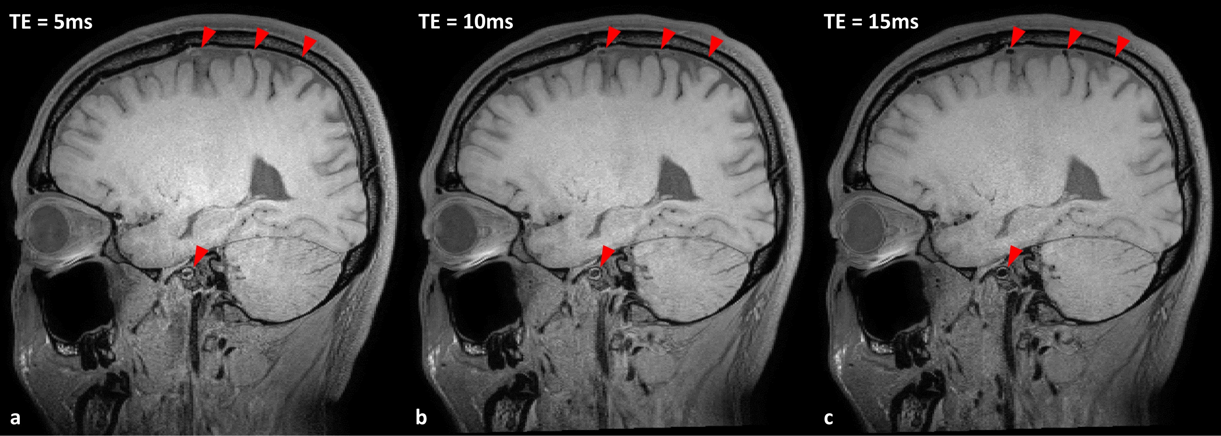
MR experiments: The optimized T1 weighted 3D TSE whole brain intracranial VWI datasets were acquired on a Philips Ingenia 3.0T MR scanner using a 32-channel head coil with FOV=230x230x210mm3, isotropic 0.5mm resolution, TR=800ms, echo spacing=5ms, adiabatic spectral fat suppression, NSA=1, x6 SENSE acceleration. With different first several skipped echoes, TE can correspondingly vary from 5ms to 15ms. Five healthy volunteers were scanned to optimize and evaluate the performance of the developed data acquisition and processing scheme.
Results
Comparison of flow suppression in small vessels: Figure 3 demonstrates that the flow suppression can be improved by skipping the first several echoes, in particular for certain segments of the internal carotid arteries and intracranial small vessels. However, increasing the number of skipped echoes may also increase TE and reduce SNR and the T1 contrast. Skipping the first two echoes provides a reasonable compromise across flow suppression, SNR and TE.
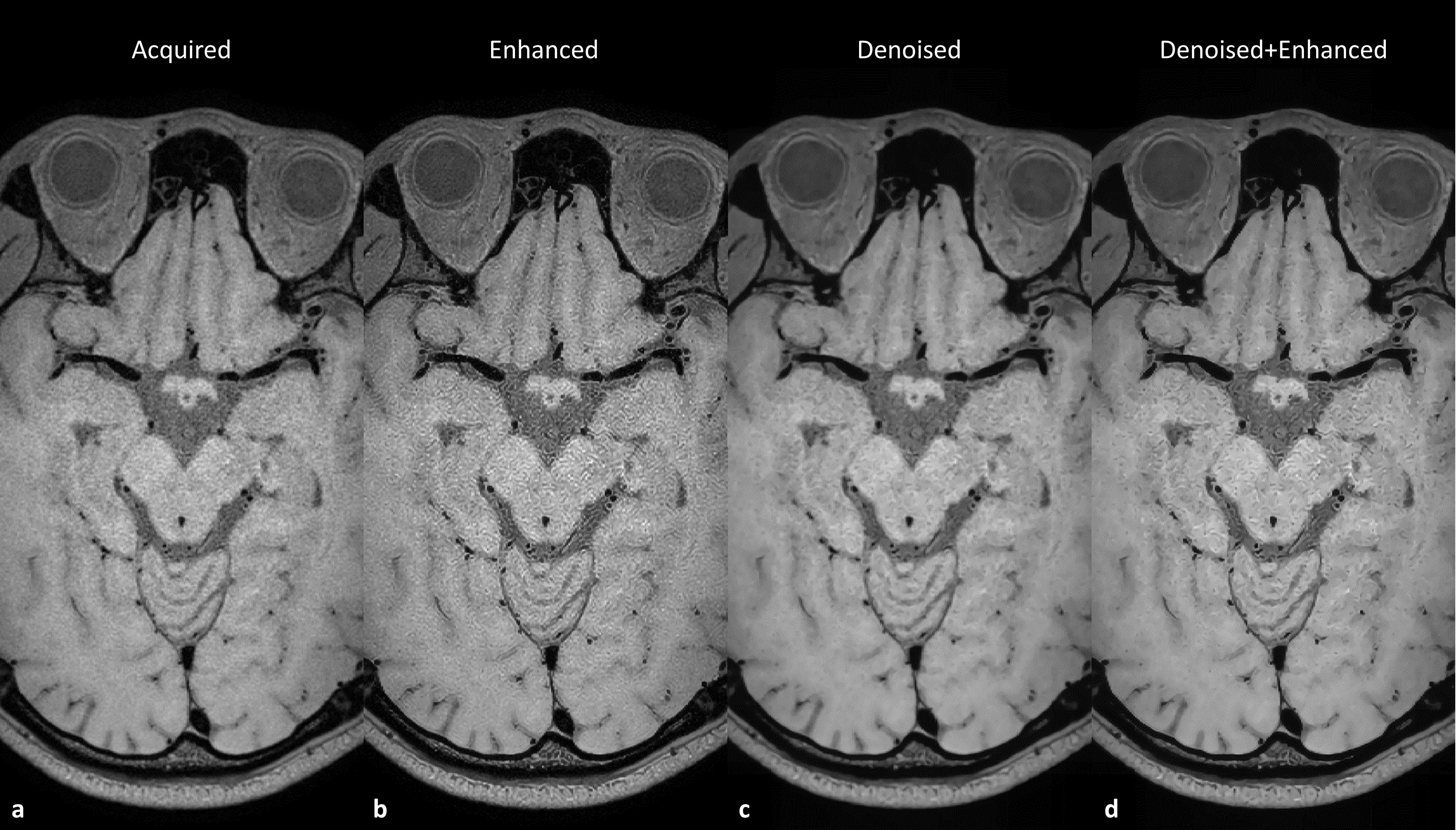
Evaluation of noise reduction and super resolution: An image quality comparison across different post-processing schemes is provided at the middle cerebral artery slice in figure 4 and basilar artery slice in figure 5. The nonlocal denoised image can effectively reduce the background noise in the originally acquired image particularly within the cerebrospinal fluid region while preserving the vessel wall boundaries. With the denoised image as input for super resolution enhancement, the sharp delineation of intracranial vessel wall boundaries was maintained but the noise artifact amplification was reduced.
Discussion and Conclusion
A T1 weighted 3D TSE sequence was optimized in terms of overall image quality and scan efficiency to allow for whole brain isotropic 0.5mm resolution intracranial VWI at 3T in 8mins28secs. The intra-voxel dephasing based flow suppression was improved for small arteries with extended echo time, but no significant change of T1 contrast was observed. The image quality is further improved by using a proper nonlocal denoising method to reduce the background noise level while preserving vessel wall boundary delineation and a super resolution method to enhance the boundary definition. This optimized imaging and post-processing technique has demonstrated promising results and may enable whole brain investigation of culprit plaques in ischemic stroke patients at 3.0T.Acknowledgements
This study is supported by the grant from National Institutes of Health (5R01NS092207). Also, we would like to acknowledge Nvidia for supporting the GPU device and the public MR IXI database (http://brain-development.org/ixi-dataset/) for training deep learning models.References
1. Qiao Y, Steinman DA, Qin Q, Etesami M, Schär M, Astor BC, Wasserman BA. Intracranial arterial wall imaging using three-dimensional high isotropic resolution black blood MRI at 3.0 Tesla. J Magn Reson Imaging. 2011;34(1):22-30.
2. Qiao Y, Steinman DA, Qin Q, Etesami M, Schär M, Astor BC, Wasserman BA. Intracranial plaque enhancement in patients with cerebrovascular events on high-spatial-resolution MR images. Radiology. 2014;271(2):534-42.
3. Yang H, Zhang X, Qin Q, Liu L, Wasserman BA, Qiao Y. Improved cerebrospinal fluid suppression for intracranial vessel wall MRI. J Magn Reson Imaging. 2016;44(3):665-72.
4. Fan Z, Yang Q, Deng Z, Li Y, Bi X, Song S, Li D. Whole-brain intracranial vessel wall imaging at 3 Tesla using cerebrospinal fluid-attenuated T1-weighted 3D turbo spin echo. Magn Reson Med. 2017;77(3):1142-1150.
5. Yang Q, Deng Z, Bi X, Song SS, Schlick KH, Gonzalez NR, Li D, Fan Z. Whole-brain vessel wall MRI: A parameter tune-up solution to improve the scan efficiency of three-dimensional variable flip-angle turbo spin-echo. J Magn Reson Imaging. 2017;46(3):751-757.
6. Zhou Z, Chen S, Wu J, Zhao X, Börnert P, Yuan C. Deep Convolutional Neural Network Enhanced 3D High Resolution Turbo Spin Echo Intracranial Vessel Wall Imaging. ISMRM 2018. p1049.
7. Wang X, Girshick R, Gupta A, He K. Non-local Neural Networks. IEEE Conference on Computer Vision and Pattern Recognition (CVPR) 2018, pp. 7794-7803.
8. Buades A, Coll B, Morel JM. A non-local algorithm for image denoising. IEEE Conference on Computer Vision and Pattern Recognition (CVPR) 2005, pp. 60-65.
Figures