3234
MRI Correlates of Neurological Outcomes in a Rat Spinal Cord Injury Model1Neurosurgery, Medical College of Wisconsin, Milwaukee, WI, United States, 2Biomedical Engineering, Marquette University & Medical College of Wisconsin, Milwaukee, WI, United States
Synopsis
This work evaluated the prognostic potential for quantitative MRI measures of the spinal cord compared to neurological assessments in a rat model of spinal cord injury. The results demonstrate in the acute setting, that a double diffusion encoded spectroscopy acquisition has greater accuracy than either DWI-EPI or T2 mapping while in the chronic setting, measures of spinal cord atrophy perform better than DWI measures. These results set the basis for future patient studies to improve MRI biomarkers in spinal cord injury.
Introduction
T2-weighted MRI is the primary diagnostic standard for spinal cord injury (SCI), but it is only a modest predictor of neurological outcome. Likewise, atrophy of the spinal cord is often associated with chronic neurological status1, but animal models have demonstrated axonal sparing is more closely tied to long-term function2. In this work, we evaluated the potential for independent or combined MRI measures to more accurately predict neurological outcome in a rat model of spinal cord contusion injury.Magnetic Resonance Imaging
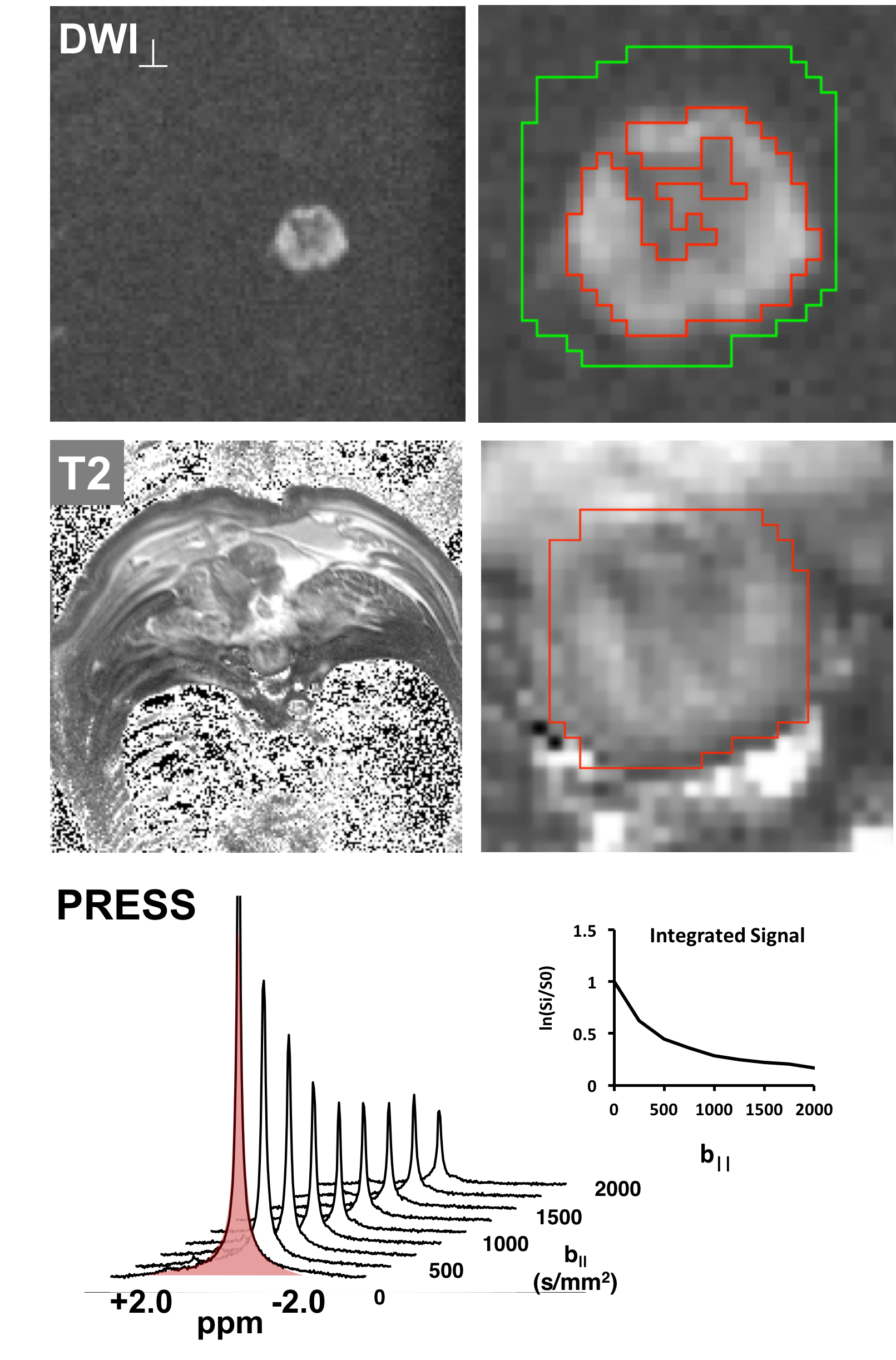
40 female Sprague-Dawley rats received a thoracic (T10) spinal cord contusion injury with the NYU impactor at varying severities (mild=9; moderate=11; severe=11; sham=9). Animals underwent MRI at 24 hours, 30 days, and 90 days post injury at the lesion site on a Bruker 9.4T MRI system using a four-channel surface Rx coil. A series of 1.5 mm thick T2-weighted axial images were acquired centered at the lesion with echo times of 17, 50, and 83 ms (TR=6000ms). Diffusion weighted images were acquired using an echo planar readout at the same locations (TR=1600ms, respiratory gated; TE=32ms; Scan time=~15 mins) using a non-traditional, cord-optimized single diffusion encoding scheme3 with vectors oriented perpendicular to the spinal cord (b-value: 0, 200, 500, 1000, & 2000), and parallel to the cord (b-value: 0, 250, 500, 750, & 1000; all with a perpendicular ‘filter’ of b=2000). A separate double diffusion encoded single-voxel spectroscopy voxel (DDE-PRESS) was also obtained at the injury epicenter (TR=3000s; TE=38ms) using 10 parallel b-values (0-1000 s/mm2) and ‘filter’ b-value of 2000 (Scan time=~2 mins).Data Analysis
Quantitative T2 maps were derived and manual regions of interest (ROIs) delineated the spinal cord. The ROIs were thresholded by T2 values to further exclude CSF (>130 ms) and hemorrhage (<30 ms) base on empirical characterization. The cord cross-sectional volume (mm3) and T2 (ms) values were obtained for subsequent analysis. ROIs for DWI were also manually delineated and thresholded by the perpendicular-weighted images with SNR values above 20, which reveal the residual cord tissue. A kurtosis model4 was used for voxelwise estimates of axonal water fraction (AWF). DW images parallel to the cord were fit to a monoexponential equation to derive what can be considered the intra-axonal diffusion, Daxial since the extracellular signals are highly suppressed by the perpendicular diffusion filter. DDE-PRESS spectra were quantified automatically by integration of the absolute-valued water peak between -2 and 2 ppm and signals were fit to a monoexponential model utilizing only the “filtered” diffusion acquisitions. Pearson’s product moment correlations and multivariate linear regression were employed to determine associations between the MRI metrics and neurological functional scores.Results
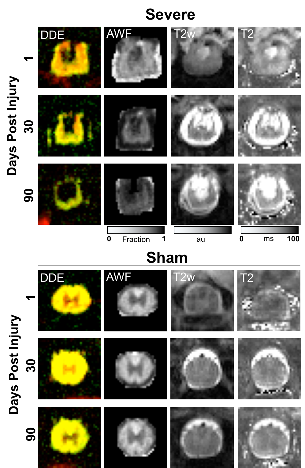
Acutely, the injured spinal cord exhibited prominent morphological changes and varying degrees of edema and hemorrhage. Daxial maps qualitatively revealed a consistent pattern of injury without CSF partial-volume effects whereas neither axonal fraction(AWF), nor cord volume were noticeably altered at the acute timepoint, as expected. Over time, these same contrasts exhibited expected trends of decreased volume and AWF whereas Daxial partially renormalized in the residual tissue.
In the quantitative analysis of injured-only animals with shams excluded (Fig 3), Daxial from DDE-PRESS was significantly correlated with BBB score at 1 day post injury. While the same Daxial metric derived from EPI was markedly less correlated with BBB score, the two metrics were correlated with one another(R2=0.30,p=0.002). Quantitative T2 values were also correlated with BBB score. It is noted that many metrics were significantly different between the injured and sham animals (not shown).
At the chronic 90-day post-injury timepoint, Daxial, hemorrhage volume, and tissue volume were all significantly correlated with BBB score. In the linear regression analysis, only tissue volume(t=3.00,p<0.008) was a significant independent predictor of BBB score across the injured animals. With sham animals included, both tissue volume and Daxial were predictors of BBB score.
Discussion and Conclusions
The results overall demonstrate two important considerations for translation to patients. First, a diffusion-weighted single voxel acquisition centered over the injury site at the acute setting provided the highest prognostic value. The improved performance over an EPI-based readout is likely related to the increased accuracy that comes with automated analysis since quantification of regions of interest in the injured cord is complicated by lower SNR, image distortions, and the presence of hemorrhage. Second, in the chronic setting, tissue volume was a much stronger predictor of chronic neurological function than diffusion metrics. This may also be related to its improved image quality and SNR compared to maps of AWF along with similar issues of accurate quantification. Collectively, these results set the basis for future patient studies to improve MRI biomarkers in spinal cord injury.Acknowledgements
We thank Kyle Stehlik, Seung Yi Lee, and Matt Runquist for experimental assistance. We are grateful for funding support from the Department of Veterans Affairs and the Bryon Riesch Paralysis Foundation.References
1-Ziegler, et al. Neurology. 2018; 2-Nashmi, Fehlings. Brain Res Rev. 2001; 3-Skinner et al. Ann Neurol. 2018; 4-Fieremans, E., et al. NeuroImage 2011.
Figures