3230
Voxelwise and morphometric analysis using diffusion tensor MRI microscopy reveals distinct microstructural and morphometric abnormalities in ferret brain development after gestational infection with the Zika virus1NIBIB, NIH, Bethesda, MD, United States, 2Henry M. Jackson Foundation, Inc, Bethesda, MD, United States, 3APG, USUHS, Bethesda, MD, United States
Synopsis
An efficient pipeline for the processing and analysis of diffusion MRI microscopy data has been applied to study neurodevelopmental abnormaities in a ferret model of Zika infection. Individual and group differences in DTI values were found using Z-score and Cohen’s D maps to compare Zika treated and untreated P0 ferret brain specimens. Morphometric abnormalities were also identified using DTI-driven tensor based morphometry (DTBM) to show reduced local volume in the developing cortex. These results highlight the utility of this pipeline and advance the basic understanding of neurodevelopmental abnormalities that can result from exposure to the Zika virus during gestation.
Introduction
The orchestration of prenatal brain development is remarkable in organization and complexity and also vulnerable to genetic mutations and adverse factors of the in-utero environment. Characterization of anatomic and cellular outcomes in neurodevelopmental disorders is generally performed in animal models and often relies on histologic evidence and coarse measurements of length and weight. Non-invasive imaging can provide more sophisticated tools for the evaluation of morphometric and microstructural abnormalities across the whole brain that may improve the understanding of these disorders. In particular, diffusion tensor MRI (DTI) microscopy can resolve small anatomic regions such as the cortical proliferation and migration zones and also is sensitive to features of the microscale tissue environment that may be altered by abnormal development(1). Furthermore, the advancement of voxelwise and DTI-driven tensor based morphometry (DTBM)(2) has enabled streamlined and sophisticated analysis of microstructure and morphometry across brain specimens. Recently, maternal infection by the Zika virus has been associated with offspring cerebral microcephaly in humans(3, 4). Although the mechanisms for this relationship are not yet well understood, studies in animal models have begun to provide important clues along several lines of research(5, 6). To extend these findings especially for the evaluation of cerebral anatomic outcomes, the ferret has been put forward as an advantageous species as it is well suited for developmental studies by its folded cortex and the timing of birth relative to gestation(7, 8). In the present study we have optimized and applied DTI microscopy analysis tools for voxelwise comparison and DTBM to evaluate the developmental effects of gestational Zika infection on neuroanatomic and microstructural outcomes in ferret brains.Methods
Ex-vivo brain specimens (n=6) were obtained from ferret kits at post-natal day 0 from litters for which the ferret jil was either infected by the Zika virus or not. The brains were imaged using a Bruker 7T microimaging system to acquire multi-shell diffusion weighted images (DWIs) with b=100-10,000 and 100 micron isotropic resolution using a 3DEPI pulse sequence. After DWI corrections and diffusion tensor(DT) fitting(9, 10), a study-specific template was generated using DTs from the untreated brains. Then each DT volume in the study was registered to the template using DT based affine and non-linear registration(11). A single brain mask was generated in template space and warped to the native space of each brain to determine the brain volume. Voxelwise Z-score and Cohen’s D effect size maps were generated to compare the treated individual and group values for fractional anisotropy (FA) and Trace (TR) with the untreated group. For morphometric analysis, maps of the Log of the Jacobian of the determinant of the deformation fields (LogJ) were calculated and also used to generate effect size maps.Results
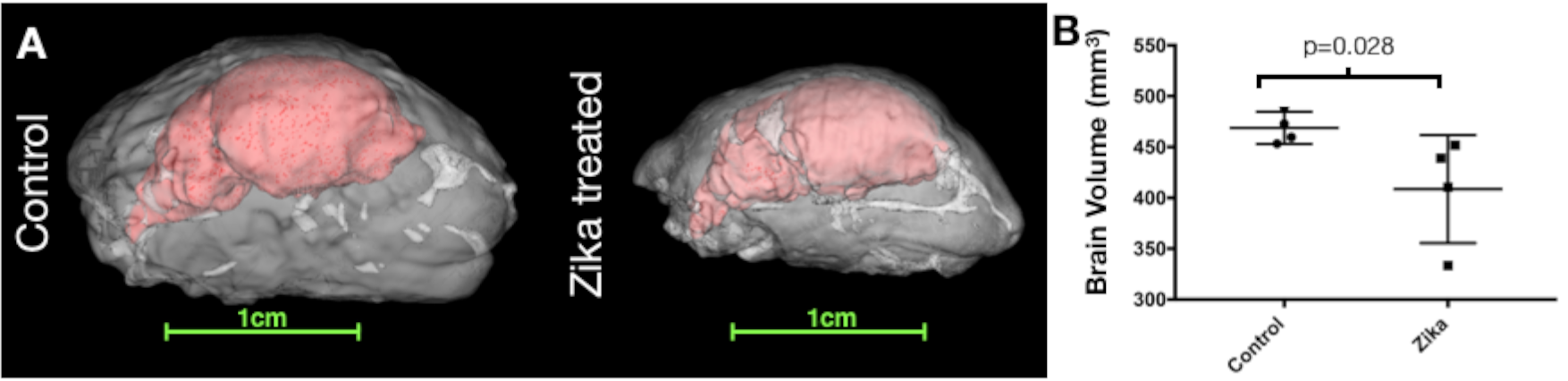
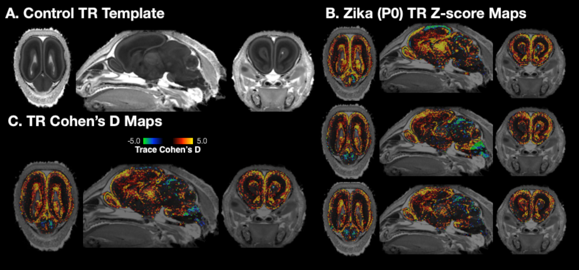
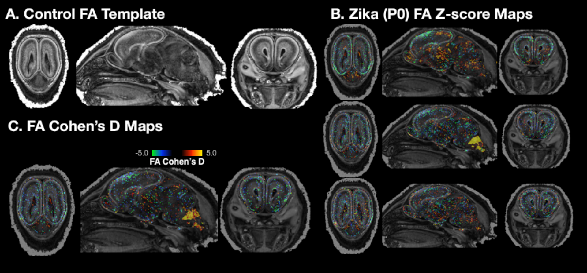
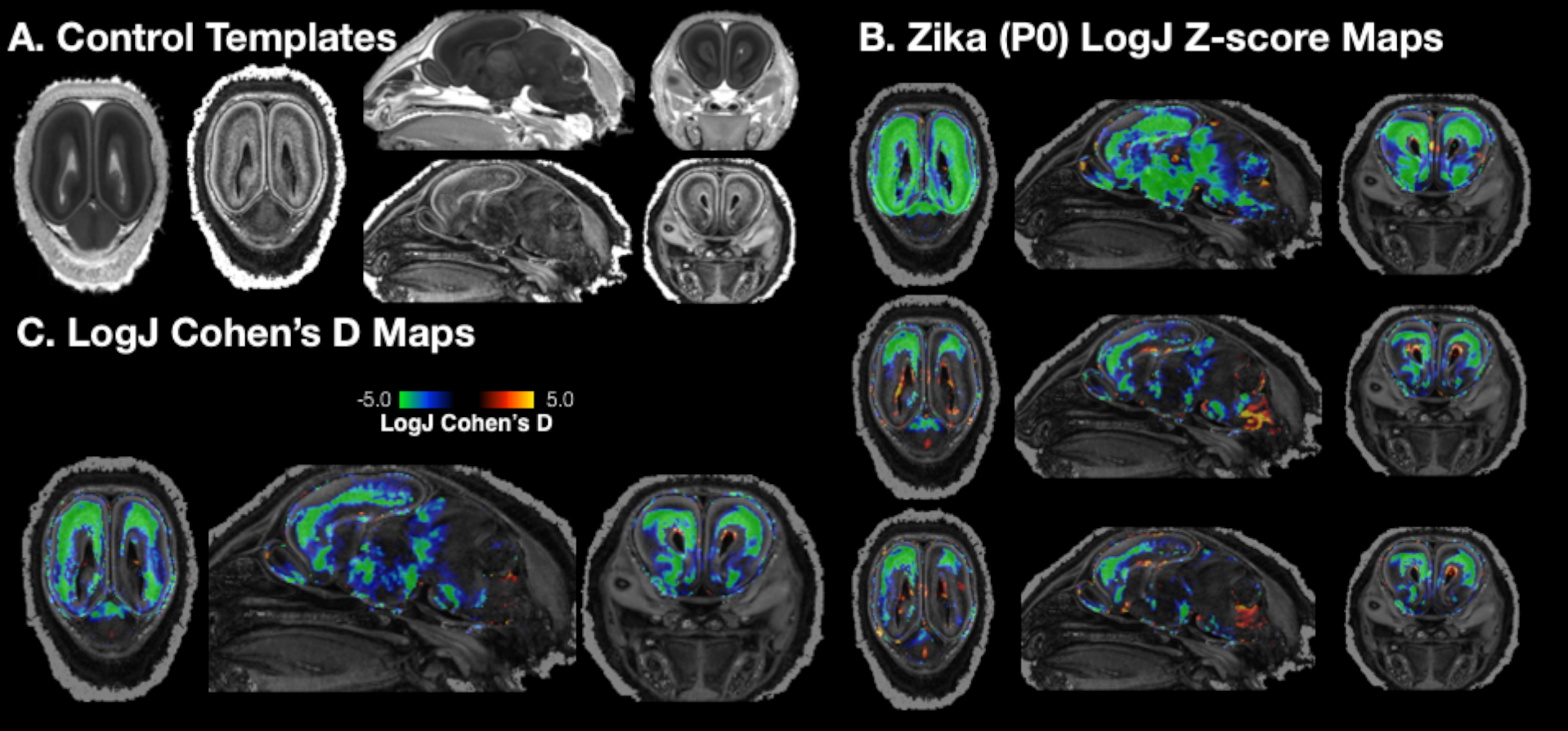
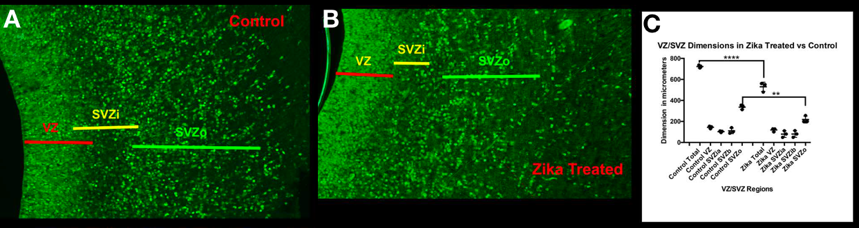
Whole brain volumes were significantly reduced in the Zika treated group (Figure 1, p=0.028 by Mann-Whitney U-test). Increased Trace values were found in the outermost cortical plate zone of the cortex by for Cohen’s D maps for group effect size and also for individual Z-score maps (Figure 2). No consistent effects were found for FA in these maps (Figure 3). The most prominent abnormality of this study was decreased local volume found in the Cohen’s D and individual Z-score maps in the ventricular and migratory zones of the cortex (Figure 4). This finding was consistent with histology from the same litters showing reduced thickness of the subventricular zone (Figure 5).Discussion
The high quality and high spatial resolution of the acquired DWI data enabled the evaluation of small anatomic regions including cortical proliferation and migration zones. The advanced registration algorithms used in this study were able to provide minimally smoothed template maps with clear tissue boundaries and anatomically faithful registration of brain structures across specimens in the study. This combination of high quality DWIs and high quality registration enabled the bias-free comparison of DTI values in treated and untreated brains ultimately resulting in novel observations about the regions affted by Zika treatment and the nature of microstructural and moprphometric outcomes namely increased diffusivity in the cortical plate and decreased local volume in other cortical zones. The similarity of Z-score and Cohen’s D maps in this study imply that the observed abnormalities are consistent across treated specimens.Conclusion
The results of this study point to specific neuroanatomic and microstructural abnormalities resulting from Zika virus treatment during gestation. The methods of this study comprise an efficient pipeline for acquisition, processing, registration and voxelwise analysis that uniquely provide combined DTI microscopy and DTBM analysis in way that is powerful for phenotyping and screening of neurodevelopmental abnormalities and other brain disorders.Acknowledgements
The authors thank Mitali Chatterjee, Francis Djankpa, William G. Valiant, Bernard Dardzinsky and Joseph J. Mattapalli for their contributions to related aspects of this work associated with the study of Zika virus in ferrets. We also thank the section for quanitative imaging and tissue science in the NICHD/NIH for enabling MRI scanning of the specimens.References
1. Huang H, Yamamoto A, Hossain M, Younes L, & Mori S (2008) Quantitative cortical mapping of fractional anisotropy in developing rat brains. The Journal of Neuroscience 28(6):1427-1433.
2. Sadeghi N, et al. (2018) Tensor-based morphometry using scalar and directional information of diffusion tensor MRI data (DTBM): Application to hereditary spastic paraplegia. Hum Brain Mapp.
3. Brasil P, et al. (2016) Zika virus infection in pregnant women in Rio de Janeiro. New England Journal of Medicine 375(24):2321-2334.
4. Moore CA, et al. (2017) Characterizing the Pattern of Anomalies in Congenital Zika Syndrome for Pediatric Clinicians. JAMA pediatrics 171(3):288-295.
5. Garcez PP, et al. (2018) Zika virus impairs the development of blood vessels in a mouse model of congenital infection. Scientific reports 8(1):12774.
6. Li H, Saucedo-Cuevas L, Shresta S, & Gleeson JG (2016) The Neurobiology of Zika Virus. Neuron 92(5):949-958.
7. Empie K, Rangarajan V, & Juul SE (2015) Is the ferret a suitable species for studying perinatal brain injury? International journal of developmental neuroscience : the official journal of the International Society for Developmental Neuroscience 45:2-10.
8. Poluch S, Jablonska B, & Juliano SL (2008) Alteration of interneuron migration in a ferret model of cortical dysplasia. Cerebral cortex (New York, N.Y. : 1991) 18(1):78-92.
9. Irfanoglu MO, Nayak A, Jenkins J, & Pierpaoli C (2017) TORTOISE v3: Improvements and New Features of the NIH Di. 25th Annual Meeting of the International Society fro Magnetic Resonance in Medicine.
10. Pierpaoli CW, L.; Irfanoglu, M.; Barnett, A.; Chang, L.-C.; Koay, C.; Pajevic, S.; Rohde, G.; Sarlls, J.; Wu, M. (2010) TORTOISE: an integrated software package for processing of diffusion MRI data. in ISMRM 18th annual meeting (Stockholm, Sweeden).
11. Irfanoglu MO, et al. (2016) DR-TAMAS: Diffeomorphic Registration for Tensor Accurate Alignment of Anatomical Structures. NeuroImage 132:439-454.
Figures