3229
Neurochemical and structural early changes and onset time in 5xFAD model: MRS and VBM studies1Korea Drug Development Platform using Radio-isotope, Korea Institute of Radiological & Medical Sciences, Seoul, Korea, Republic of, 2Division of Applied RI, Korea Institute of Radiological & Medical Sciences, Seoul, Korea, Republic of
Synopsis
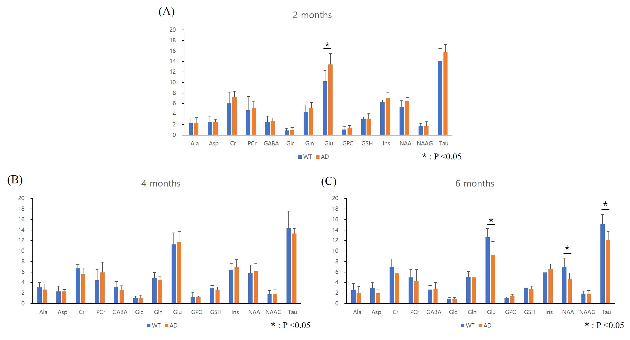
We investigated early neurochemical and structural changes in 5xFAD model using magnetic resonance spectroscopy (MRS) and Voxel-based morphometry (VBM). At 6 months, it was confirmed that Glu, NAA, Tau and volume, which is characteristic of Alzheimer’s disease (AD) model, decreased in hippocampus of 5xFAD model.
Introduction
5xFAD mouse model is useful for understanding AD mechanism and for therapy studying. This model is characterized by very aggressive amyloid deposits. Aβ42 is expressed in the brain at 1.5 months and the plaque develops at 2 months. At 4 months, the memory declines and then the neuron loss at 9 months.1 VBM and proton magnetic resonance spectroscopy (1H-MRS) are good tool for understanding the pathogenesis and the diagnosis of AD.2 In the current study, we investigated neurochemical and structural changes and the time of onset in the hippocampus of the 5xFAD model using MRS and VBM.Subjects and Methods
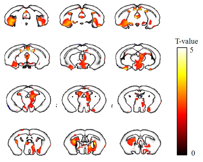
5xFAD mice group (male, 𝑛 = 6) and wildtype mice group (male, 𝑛 = 6) were used for this study. MRS were obtained at 2, 4, and 6 months of age and T2w image for VBM about mice were obtained at 6 months. All scans were performed on an Agilent 9.4T VNMRS 30cm horizontal -bore system (Agilent Inc, Palo Alto, CA, USA) with 27mm birdcage radiofrequency (RF) coil and a quadrature mouse brain surface coil (RAPID, Germany). To confirm metabolite change, 1H-MRS data was obtained using a Point-Resolved Spin echo Sequence (PRESS) with TR = 5000 ms, TE1 = 7.66 ms, TE2 = 6.01 ms, 512 averages, 8 dummy scans, 2048 acquisition data points, spectral width 5000 Hz and VAPOR (variable power and optimized relaxation delays) water suppression. A single voxel of 1.2 ×1.5 × 2 mm3 was centered on the left hippocampus for MRS position. Metabolite concentrations were analyzed by using the linear combination of model spectra (LCModel) with water signal as a reference. A SPSS was used for statistical analysis. To confirm structure change, the 3D T2-weight image was obtained fast spin each 3D sequence as follows: TR = 2500 ms, TE = 7.5 ms, TI =400ms, ETL=6, NEX=1, slab=10mm, FOV=20x20x10mm, Matrix = 128x 128x 64. For VBM analysis, SPM12 with SPMMouse tool was used for statistical analysis and preprocessing include segmentation, modulation, normalization, and smoothing. The difference of brain volume between 5xFAD mice and wildtype mice were analyzed by two-sample t-test (p<0.05, p-uncorrected).Results and Discussion
At 2 months, there was a difference between the two groups only in glutamate (Glu), and the 5xFAD mice group was higher than the wildtype mice group (P=0.022, Figure1A). At 4 months, there was no difference in any metabolic (Figure1B). However, at 6 months, there were statistical difference in Glu, N-acetylaspartate (NAA) and taurine (Tau) (p=0.021, p=0.019 and p=0.01, respectively). It was confirmed that the 5xFAD mice group was lower than the wildtype mice group in all of three metabolic (Figure1C). A decrease in Glu and NAA characteristic of AD occurred at 6 months in 5xFAD.3 In addition, it showed a decrease in Tau similar to a transgenic mouse carrying the P301L mutation.4 In the VBM results, 5xFAD mice group tended to decrease in size of most brain regions than wildtype mice group at 6 months. In addition, we confirmed that the size of the hippocampus region which is characteristic of AD was reduced at 5xFAD (Figure 2). Based on our findings, the present study suggests that 5xFAD mice showed the characteristics of AD from the result of MRS and VBM and confirmed the change of brain from MRI at least 6 months.Conclusion
We found that a decrease in Glu, NAA, Tau and volume in the hippocampus of 5xFAD mouse model from 6 months.Acknowledgements
This work was supported by the National Research Foundation of Korea (NRF) grant funded by the Korea government (Ministry of Science, ICT) (NRF-2013M2C2A1074238)References
1. Oakley, H. et al. Intraneuronal beta-Amyloid Aggregates, Neurodegeneration, and Neuron Loss in Transgenic Mice with Five Familial Alzheimer’s Disease Mutations: Potential Factors in Amyloid Plaque Formation. J. Neurosci. (2006)
2. Liang, S. et al. Magnetic resonance spectroscopy analysis of neurochemical changes in the atrophic hippocampus of APP/PS1 transgenic mice. Behavioural brain research, (2017).335, 26-31.3
3. Choi. JK. et al. Application of MRS to mouse models of neurodegenerative illness. NMR Biomed (2007) 20, 216-237
4. Santacruz. K. et al. Tau suppression in a neurodegenerative mouse model improves memory function. Science (2005) 309, 476-481
Figures