3228
Evaluation of Diffusion-Weighted Imaging of the Macaque Brains Using Diffusion-Prepared TSE1Interdisciplinary Institute of Neuroscience and Technology, Qiushi Academy for Advanced Studies, College of Biomedical Engineering & Instrument Science, Zhejiang University, Hangzhou, China, 2Center for Magnetic Resonance Research, School of Medicine, University of Minnesota, Minneapolis, MN, United States
Synopsis
A major challenge with EPI-based
diffusion-weighted imaging (dMRI) is magnetic field inhomogeneity-associated
distortion and signal loss. We implemented a mono-polar diffusion-preparation
module for TSE sequence (DP-TSE) as an alternative to achieve distortion-free,
high-resolution diffusion imaging with improved signal-to-noise ratio (SNR).
Such an approach has been demonstrated in human subjects with a promising
potential. We want to further evaluate the robustness of the implemented DP-TSE
sequence and the feasibility of applying this approach for anesthetized macaques,
and investigate whether DP-TSE is superior to alternative dMRI method in terms
of imaging quality and SNR efficiency.
Purpose
Diffusion weighted MRI (dMRI) has been widely used in both neuroscience research (as exemplified by the Human Connectome Project) and clinical applications as an essential tool of investigating connectivity and tissue microstructure 1-3. A major challenge with EPI-based diffusion-weighted imaging (dMRI) is magnetic field inhomogeneity-associated distortion and signal loss. We implemented a mono-polar diffusion-preparation module for TSE sequence (DP-TSE) as an alternative to achieve distortion-free, high-resolution diffusion imaging with improved signal-to-noise ratio (SNR). Such an approach has been demonstrated in human subjects with a promising potential . In the present study, based upon our preliminary work4, we have implemented and evaluated a TSE-based method for dMRI in an anesthetized macaque, in which diffusion-preparation module was conjugated to a TSE readout scheme (refrred to as Diffusion-Prepared TSE, DP-TSE); a prototype readout segmented EPI (rsEPI) sequence was also applied for comparison. The study results are reported as below.Methods
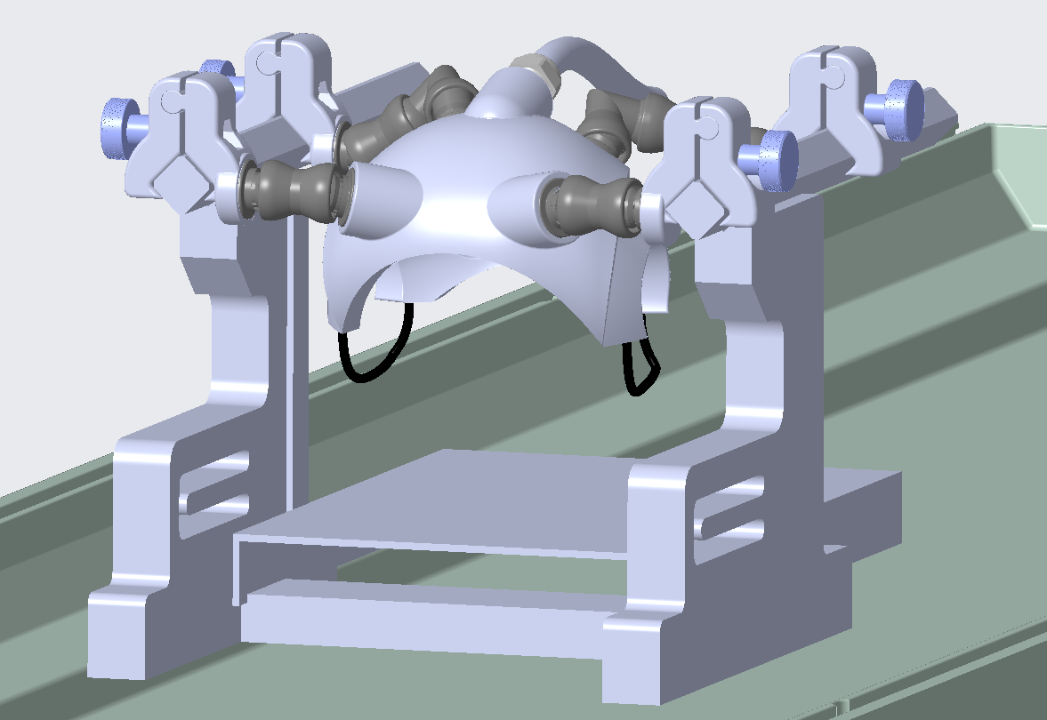
The studies were performed on a Siemens 3T Prisma MRI scanner equipped with a customized 16-channel helmet-like monkey head array fixed over the stereotaxic frames5 as shown in Figure 1. Images were acquired from a female macaque (13-year old, 9.6kg) that was anesthetized and maintained by 1.4% to 2% isoflurane. All procedures were in accordance with NIH standards and approved by our Institutional Animal Care Committee. During imaging acquisition, the macaque was tightly placed in the sphinx position with its head centered within the head coil.
To evaluate the robustness and time efficiency of the TSE-based method for in vivo macaque diffusion imaging, we performed full-brain macaque dMRI with the DP-TSE and the rsEPI sequences6. Parallel imaging with GRAPPA and relatively high b-values were used for comparison. Imaging parameters were as follows: FOV = 160 x 160 (rsEPI) or 128 x 128 (DP-TSE) mm2; matrix size = 160 x 160 (rsEPI) or 128 x 128 (DP-TSE); in-plane resolution = 1 x 1 mm2; slice thickness = 2 mm; 24 slices; b-value = 500, 1,000, 1,500, 2,000 s/mm2; DP-TSE: turbo factor = 11, TE = 11 ms; rsEPI segments = 7. In one slice ADC fitting TR = 1500 ms and whole brain DWI scan reEPI TE and both TRs were optimized as short as possible with TR = 4000- 4500 ms and rsEPI TE = 80-85 ms. Whole brain three-direction DWI scan time of different protocols are lists in Table 1. Post-processing, including co-registration and reconstruction of ADC maps, were performed in MATLAB.
Results and Discussions
Our study indicated that the performance of the implemented DP-TSE is satisfactory for diffusion imaging with anesthetized macaques. With careful preparations to ensure the monkey head well stabilized, using the DP-TSE method, high-resolution and distortion-free diffusion images were successfully obtained.
The diffusion-weighted images (DWIs) and ADC maps from two protocols with b = 1,000 s/mm2 are displayed in Figure 2. ADC maps from DP-TSE with a larger b-value are presented in Figure 3(a); ADC maps using two protocols are shown in Figure 3(b); DP-TSE images with and without parallel-imaging acceleration are shown in Figure 3(c). Our study suggested that DP-TSE and rsEPI methods have similar diffusion SNR efficiency.
It is worthy noting that the DP-TSE method is superior to the rsEPI approach as indicated by the study results (Figure 3(a)); when higher b values are used, the DP-TSE method was able to provide high-quality diffusion imaging results but the rsEPI approach could not. Further more, utilizing higher parallel imaging acceleration factors can further increase the time efficiency of diffusion imaging with the DP-TSE (Figure 3(c)).
Conclusions
The implemented DP-TSE sequence can achieve distortion free, high SNR and time-efficient diffusion weighted imaging in anesthetized monkeys.Acknowledgements
National Natural Science Foundation 81701774 and 61771423, NIH NS093998. We thank Kamil Ugurbil, Christopher Kroenke, Michael Reusz, Zheng Liu, Robert Friedman and Mykyta Chernov for helpful discussion and technical support.References
- Johansen-Berg H, Behrens TE, editors. Diffusion MRI: from quantitative measurement to in vivo neuroanatomy. Academic Press; 2013 Nov 4.
- Vu AT, Auerbach E, Lenglet C, Moeller S, Sotiropoulos SN, Jbabdi S, Andersson J, Yacoub E, Ugurbil K. High resolution whole brain diffusion imaging at 7T for the Human Connectome Project. Neuroimage. 2015 Nov 15; 122:318-331.
- Song SK, Yoshino J, Le TQ, Lin SJ, Sun SW, Cross AH, Armstrong RC. Demyelination increases radial diffusivity in corpus callosum of mouse brain. Neuroimage. 2005 May 15; 26(1):132-40.
- Zhang J, Li X, Ugurbil K, Roe AW, Zhang X, and Wang D. Evaluation of Monopolar Diffusion-Prepared TSE for Diffusion Imaging. Intl. Soc. Mag. Reson. Med. 26 (2018): 1619.
- Gao Y, Wang X, Friedman R, Chernov M, Kroenke C, Roe AW, and Zhang X. 16-Channel Array Coil for Anesthetized Monkey Multi-modal Neuroimaging at 3T. Submitted to ISMRM 2019.
- Wang P, Zhang J, Qian M, Sun Y, Wang D, and Zhang X. Evaluation of Diffusion-Weighted Imaging of the Macaque Brains Using Readout-Segmented EPI at 7T. Intl. Soc. Mag. Reson. Med. 26 (2018): 3306.
Figures