3226
Neuroimaging of Nipah Virus infection in an African Green Monkey Model1IRF, NIAID/NIH, Frederick, MD, United States, 2Clinical Research Directorate/Clinical Monitoring Research Program, Leidos Biomedical Research, NCI Campus, Frederick, MD, United States, 3Virus Ecology Unit, Laboratory of Virology, Rocky, Mountain Laboratories, NIAID/NIH, Hamilton, MT, United States, 4Clinical Center, NIH, Bethesda, MD, United States
Synopsis
The purpose of this study was to utilize MRI to assess alterations in the brain that occur in African Green Monkeys infected with Nipah virus (NiV) via aerosol inoculation. Within 15 days of exposure to NiV, signal alterations were observed in the brain in T2-weighted, fluid-attenuated inversion recovery, and diffusion-weighted images, suggestive of infarction, inflammation and edema induced by NiV. The identification of non-invasive imaging biomarkers of acute NiV neurologic disease progression in this animal model could aid in the examination of potential vaccines and therapeutics.
Introduction
Nipah virus (NiV) is an emerging virus associated with outbreaks of acute respiratory disease and encephalitis in India and Bangladesh with a case fatality rate exceeding 50% [1]. The virus which was first identified and isolated in 1999, has historically been considered “encephalitic” based on Malaysian patients with neurologic disease. The brain is one of the most severely affected organs, and patients can present with fever, headache, dizziness, areflexia, hypertension, vomiting, reduced levels of consciousness and occasionally seizures. To date, the mechanisms of the disease are poorly understood and no effective vaccines or treatments have been available to human.
Animal models of NiV infection are of interest for the investigation of disease pathogenesis, correspondence to human disease, and development of medical countermeasures [2-4]. The development of NiV disease has been previously investigated in the African Green Monkey (AGM) following intratracheal or aerosol exposure using small (<3 μm) and intermediate particle (≥7 μm) inoculates with doses ranging from 100 to 10,000 plaque forming units (pfu) [3, 4]. Results from one study demonstrate that aerosol exposure to intermediate particles (>6.5 μm) at a lower challenge dose (~100 pfu) than previously used results in an extended disease course (12.5 days) relative to intratracheal or small particle aerosol exposure (8 days) [4-5]. Therefore, exposure with large particle (>10 μm) aerosol at a lower dosage is likely to extend the disease course and allow examination of delayed-onset symptoms (e.g., neurological symptoms).
The objective of this study was to utilize MRI to assess structural brain changes that occur in an AGM model of NiV infection by a lower aerosol dose of larger particle (11‒13 µm) of NiV than previously used to mimic a more protracted human disease course compared to that observed in previous animal studies.
Methods
Six adult AGMs (3‒7 kg) that had been pre-screened for NiV antibodies were exposed to a large particle (11-13 µm) aerosol challenge (~500 pfu) of NiV Malaysia strain. All animals underwent 60 minutes of imaging on a Philips Achieva 3 Tesla clinical MR scanner (Philips Healthcare, Cleveland, OH, USA). Using an 8-channel pediatric neuro-spine coil, brain images (T2-weighted, FLAIR, R2*, DTI, and T1w pre and post-contrast images in transaxial imaging planes) were obtained at baseline, every three days post-exposure (PE) for two weeks, and every week afterwards until week 8. For each scan, subjects were intubated, anesthetized using isoflurane and positioned supine on the scanner bed. Clinical scores and Nipah antibodies were measured.Results
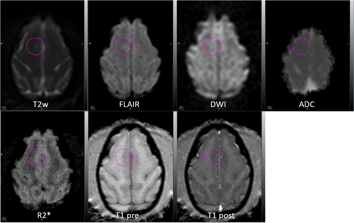
Four out of six animals showed abnormalities reminiscent of human pathology in MRI, including one animal that survived the infection. In the survivor (#8671), two focal high signal intensity lesions were seen on FLAIR and T2-weighted images in the left cerebellar and right frontal subcortical white matter (Fig. 1) starting on day 12 PE. The latter resolved on day 22. The lesions showed facilitated diffusion. In the second animal (#8078), a small focus of restricted diffusion was seen in the left frontal subcortical white matter on day 15 PE. Multiple bilateral cerebral lesions eventually developed on day 17 PE, mostly showing restricted diffusion (Fig 2). In the third animal (#8233), multiple lesions were seen on FLAIR and T2-weighted images on days 15 and 20 PE in the pons and subcortical white matter of both cerebral hemispheres. The lesions showed restricted (red) or facilitated (green) diffusion or a combination of both. In the fourth animal (not shown), only one focus of restricted diffusion was seen in the right internal capsule on day 12 and 15, with no enhancement. Three out of six animals showed Nipah-specific antibodies including one of the AGM survivors (#8671), while the other survivor (#8212) not.Discussion
Four out of 6 animals showed abnormalities on brain MRI ranging from cytotoxic edema (restricted diffusion) to vasogenic edema (facilitated diffusion) or a combination of both. The majority of the lesions we saw were small infarcts although a few probably reflect inflammatory/encephalitic changes. Resolution or decreased size of some of those lesions resembles what has been reported in patients with NiV infection. Further quantitative imaging analyses with viral load, cytokines, cerebrospinal fluid, and pathology will be performed.Conclusion
We have shown structural brain changes in an AGM model of NiV infection exposured with a lower dose of large-particle NiV aerosol (8‒12 µm) which were similar to human disease manifestations. Our animal model will help us better understand the acute neurologic features of NiV infection.Acknowledgements
Animal Ethics Statement: Animals were housed in an AAALAC-International-accredited facility. All experimental procedures were approved by the NIAID Division of Clinical Research (DCR) Animal Care and Use Committee and were in compliance with the Animal Welfare Act regulations, Public Health Service policy, and the Guide for the Care and Use of Laboratory Animals recommendations.
Funding: This work was supported by NIAID Division of Intramural Research and NIAID DCR and was performed under Battelle Memorial Institute contract (No. HHSN272200700016I) with NIAID. Additional support was provided by the NCI Contract No. HHSN261200800001E.
References
1. Kulkarni, Venkatesh, Kumar, “Nipah virus infection: current scenario”, Indian J virol 24(3): 398-408 (2013).
2. Lentz M, Hammoud D, Cong Y, et al. Neuroimaging of Nipah virus in a Syrian hamster model of infection. Presented at the International Society for Magnetic Resonance in Medicine 24th Annual Meeting, 07-13 May 2016, Singapore.
3. Johnston SC, Briese T, Bell TM, et al. Detailed analysis of the African green monkey model of Nipah virus disease. PLoS One. 2015;10(2): e0117817.
4. Cong Y, Lentz MR, Lara A, et al. Loss in lung volume and changes in the immune response demonstrate disease progression in African green monkeys infected by small-particle aerosol and intratracheal exposure to Nipah virus. PLoS Negl Trop Dis.2017;11(4): e0005532.
Figures