3224
Neuroimaging Microglia: Development of a Quantitative Multi-Compartment Diffusion MRI Biomarkers of Microglial Density1Departments of Radiology, Biomedical Engineering, and Psychiatry, University of Wisconsin School of Medicine and Public Health, Madison, WI, United States
Synopsis
Neuroinflammation plays a critical role in neurologic and psychiatric disorders of the central nervous system (CNS) from ischemic stroke and traumatic brain injury to Alzheimer’s disease, schizophrenia, and major depression. The application of multi-compartment diffusion MRI techniques for the robust, non-invasive, and quantitative evaluation of microglial morphology and density in the setting of acute and chronic neuroinflammation would represent an important advancement in understanding, identification, and therapeutic monitoring of microglia across a broad spectrum of acute and chronic disorders of the CNS. We present the first evidence and application of multi-compartment diffusion MR techniques for the sensitive detection of changes in microglial density throughout the brain.
Introduction
Brain-immune interactions contribute to numerous acute and chronic disorders of the central nervous system (CNS) from ischemic stroke and traumatic brain injury to age-related dementia and Alzheimer's disease.1,2 Schizophrenia, major depression, and other neuropsychiatric disorders also exhibit hallmarks of neuroinflammation where peripheral cellular and humoral immunological abnormalities are more prevalent in psychiatric patients than in healthy controls. Genome-wide studies in schizophrenia have also revealed an association with markers in the major histocompatibility complex region (MHC) and post-mortem studies have provided evidence of increased microglial populations and microglial activation in patients with schizophrenia, depression, and other affective disorders.3,4 With a growing recognition of the role of microglial-mediated neuroinflammation in the psychiatric disorders, there is significant interest in new methodologies aimed at imaging neuroinflammation and microglial populations. However, many current imaging methodologies are encumbered by significant limitations including low specificity (diffusion tensor imaging [DTI]), challenges to quantification (PET imaging of translocator protein [TSPO]), and low biocompatibility/toxicity (microparticles of iron oxide [MPIO]). Despite active research efforts to overcome these limitations, there remains no safe, widely accessible, or clinically viable neuroimaging methodology available for the in vivo study of neuroinflammation. We present herein the first evidence of the sensitivity and robustness of the extra-neurite compartment in multi-compartment diffusion MRI to detect alterations in microglial morphology and density throughout the brain and further characterize the relationship between quantitative measures of the extra-neurite compartment with microglial density.Methods
Microglial activation is induced in a rat model of peripheral inflammation via a single intraperitoneal (ip) injection of lipopolysaccharide (LPS). Briefly, male rats are injected with a single ip dose of LPS or vehicle (n=8, each group). 3- hours post injection, all animals are transcardially perfused with PFA and brains cleanly dissected from the cranial vault. Brains are simultaneously imaged with a 4.7-T Agilent MRI system and a 3.5-cm diameter quadrature volume RF coil was used to acquire 10 non-diffusion weighted images (b=0 s⋅mm-2) and 75 diffusion weighted images (25: b=800 s⋅mm-2, 50: b=2,000 s⋅mm-2). Following standard preprocessing, tensors are reconstructed, registered, and normalized to a population-specific template. In parallel, NODDI parameters were estimated using an ex-vivo fitting model and converted to volumetric parameter maps. Transformation fields from corresponding single-shell data registration were applied to NODDI data. NODDI data were then projected onto the previously-defined white matter skeleton that was created from single-shell diffusion tract-based spatial statistics (TBSS). TBSS were computed to evaluate whole-brain voxel-wise differences in standard diffusion indices and NODDI parameters between LPS and vehicle treated animals. Permutation test results for multiple comparisons and threshold-free cluster enhancement (TFCE) are implemented with FSL's Randomize and considered significant at the <0.05 level following family-wise error correction. Following imaging, brains were removed from their custom holders and were returned to ice-cold 4% PFA for 24 hours. After cryoprotection with 30% sucrose in 1X PBS, 40 um coronal floating sections were collected in 1X PBS and stored at 4C until staining. Immunostaining and fluorescence quantification with Iba-1, NeuN, and GFAP were then performed per protocol.Results
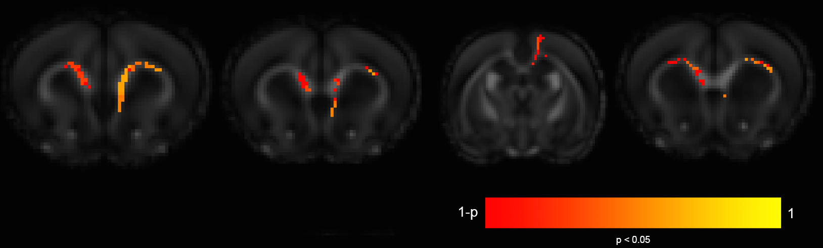
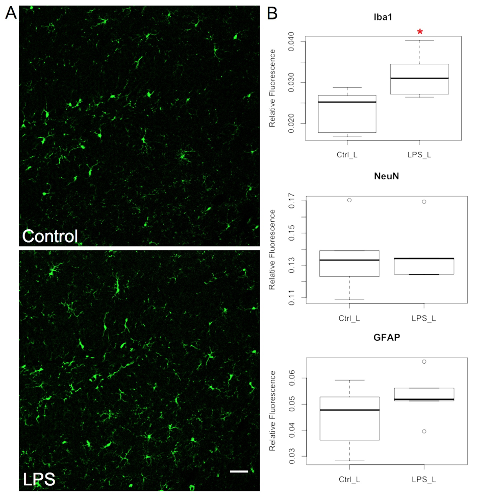
TBSS and ROI analysis reveals numerous clusters of significantly increased voxel-wise change in ODI 3-hours following intraperitoneal injection of LPS (Fig. 1). Concurrently performed quantitative immunofluorescence demonstrates significant changes in microglial populations in calculated imaging ROIs buttressing both the sensitivity and specificity of ODI to microglial populations (Fig. 2). Notably, no voxel-wise changes are seen in any scalar measures of the diffusion tensor or in parametric measures of NDI thus confirming the unique sensitivity of ODI to morphometric changes in activated microglia in model of microglial activation (Fig. 3).Conclusions
Neuroinflammation underlies numerous neurologic and neuropsychiatric disorders and noninvasive biomarkers to detect and monitor microglial inflammation are needed. Multi-compartment diffusion tensor models such as NODDI allow for the direct biophysical interrogation of neurite density and orientation but also fortuitously permit the simultaneous interrogation of the extracellular compartment and the neuropathological changes that can occur within this space including changes related to microglia-mediated neuroinflammation. We will present the potentially groundbreaking application of NODDI to measures changes in the extracellular compartment to noninvasively detect and measure microglial burden in the brain. Ongoing work will determine the quantitative relationship between microglial density with parametric measures of NODDI with quantitative immunofluorescence and stereology. If successful, the validation of NODDI to detect and monitor microglial neuroinflammation would represent a major advance across a wide spectrum of neurologic and neuropsychiatric disorders with far reaching implications in clinical diagnosis, risk stratification, and therapeutic monitoring where a NODDI biomarker of neuroinflammation could serve as a clinical endpoint assisting in the development of critically needed therapies.Acknowledgements
This work was supported by the Department of Radiology at the University of Wisconsin School of Medicine and Public Health, the Brain and Behavior Research Foundation (NARSAD) Young Investigator Grant, and a RSNA R&E Foundation Research Fellow Grant (RF #1417). Additional support was provided by the Clinical and Translational Science Award (CTSA) program, through the NIH National Center for Advancing Translational Sciences (NCATS), grant UL1TR002373. Additional imaging support was provided by the University of Wisconsin Carbone Cancer Center Support Grant P30CA014520. The content is solely the responsibility of the authors and does not necessarily represent the official views of the NIH.References
1. Woodcock T, Morganti-Kossmann MC. The role of markers of inflammation in traumatic brain injury. Front Neurol 2013;4 MAR.
2. Iadecola C, Anrather J. The immunology of stroke: From mechanisms to translation. Nat Med 2011;17:796–808.
3. Torres-Platas SG, Cruceanu C, Chen GG, et al. Evidence for increased microglial priming and macrophage recruitment in the dorsal anterior cingulate white matter of depressed suicides. Brain Behav Immun 2014;42:50–9.
4. Trépanier MO, Hopperton KE, Mizrahi R, et al. Postmortem evidence of cerebral inflammation in schizophrenia: a systematic review. Mol Psychiatry 2016;21:1009–26.
Figures